Copyright
©The Author(s) 2022.
World J Clin Cases. Jun 26, 2022; 10(18): 6009-6020
Published online Jun 26, 2022. doi: 10.12998/wjcc.v10.i18.6009
Published online Jun 26, 2022. doi: 10.12998/wjcc.v10.i18.6009
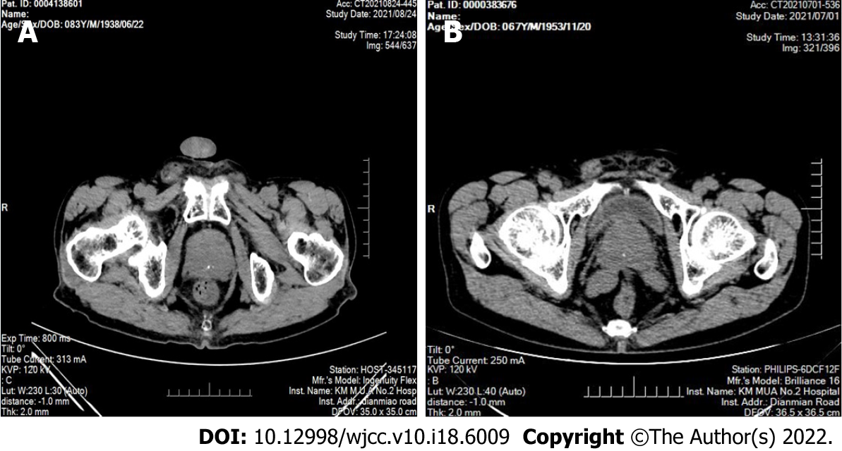
Figure 1 Computed tomography images of sclerosing adenopathy of the prostate.
A: Computed tomography (CT) showing an enlarged prostate with multiple calcifications; B: CT showing that the prostate was enlarged and calcified, partially protruding into the trigone of the bladder, and the enhancement was not uniform.
- Citation: Feng RL, Tao YP, Tan ZY, Fu S, Wang HF. Prostate sclerosing adenopathy: A clinicopathological and immunohistochemical study of twelve patients. World J Clin Cases 2022; 10(18): 6009-6020
- URL: https://www.wjgnet.com/2307-8960/full/v10/i18/6009.htm
- DOI: https://dx.doi.org/10.12998/wjcc.v10.i18.6009


