Copyright
©The Author(s) 2022.
World J Clin Cases. Jun 16, 2022; 10(17): 5884-5892
Published online Jun 16, 2022. doi: 10.12998/wjcc.v10.i17.5884
Published online Jun 16, 2022. doi: 10.12998/wjcc.v10.i17.5884
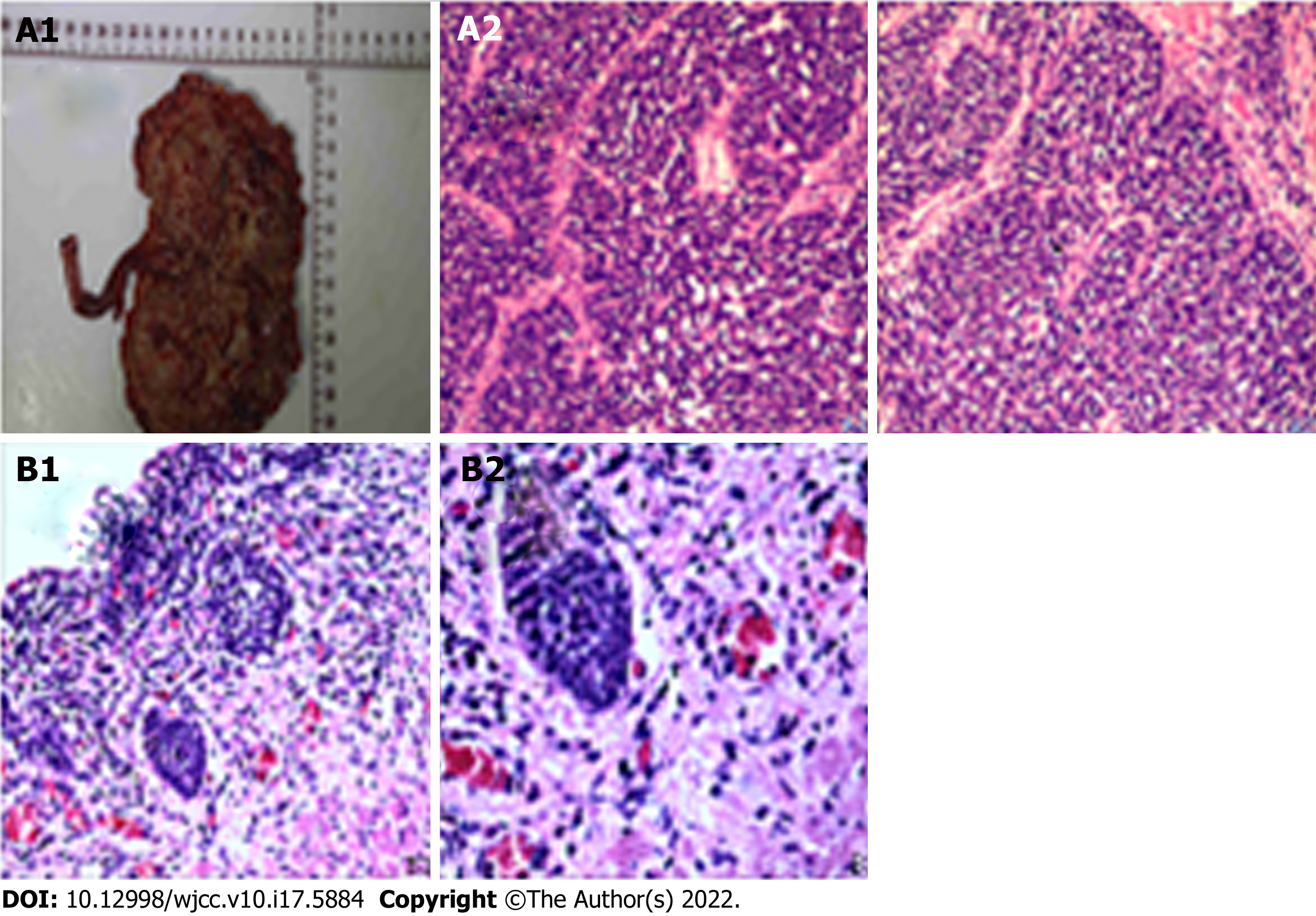
Figure 3 Postoperative pathology in 2020.
High-grade invasive papillary urothelial carcinoma and small cell carcinoma were suspected. A: Under the microscope, tumor cells were composed of two components: one type of cells was nest-like arrangement, with larger cell volume, abundant cytoplasm, large nuclei, deep staining and obvious atypia; the other type of cells was uniformly distributed in sheets, with medium to small cells, abundant cytoplasm or medium cytoplasm, and round nuclei. And the boundary between the two forms is clear (A1 and A2); B: There was no obvious abnormality at the broken end of the ureter (B1 and B2).
- Citation: Xie K, Li XY, Liao BJ, Wu SC, Chen WM. Primary renal small cell carcinoma: A case report. World J Clin Cases 2022; 10(17): 5884-5892
- URL: https://www.wjgnet.com/2307-8960/full/v10/i17/5884.htm
- DOI: https://dx.doi.org/10.12998/wjcc.v10.i17.5884


