Copyright
©The Author(s) 2022.
World J Clin Cases. Jun 16, 2022; 10(17): 5861-5868
Published online Jun 16, 2022. doi: 10.12998/wjcc.v10.i17.5861
Published online Jun 16, 2022. doi: 10.12998/wjcc.v10.i17.5861
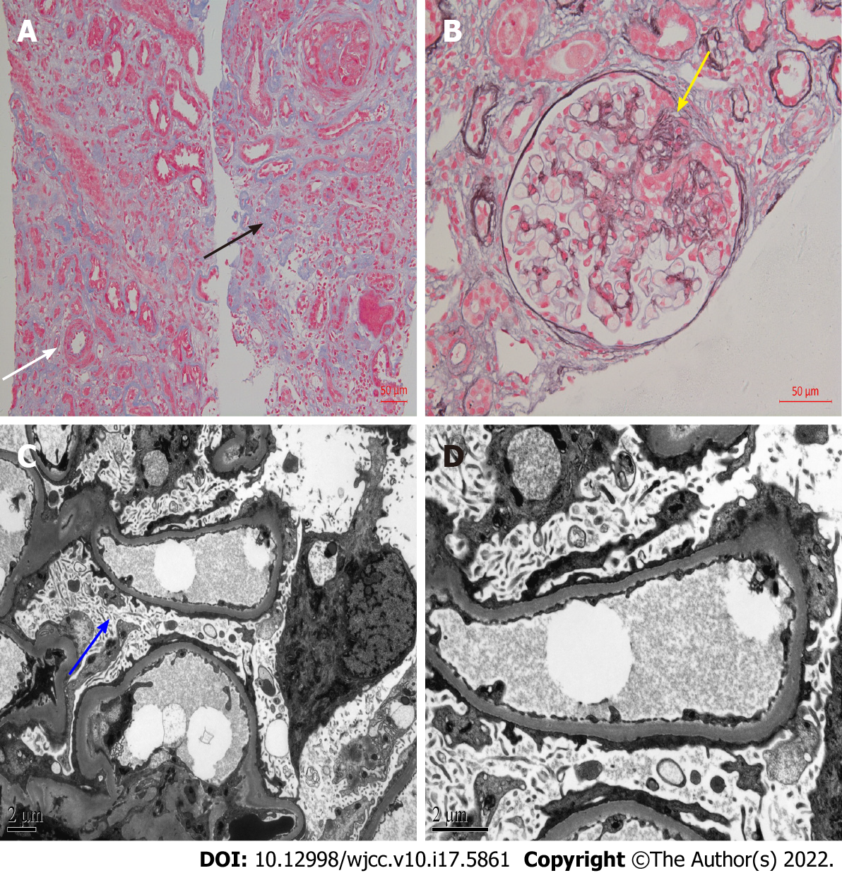
Figure 2 Light microscopy and electron microscopy of histological changes of renal biopsy after 20 years.
A: Masson, × 200. Multifocal and patchy atrophy of renal tubules, multifocal and patchy lymphocytic infiltration of renal interstitium with fibrosis (black arrow), and thickening of arterioles (white arrow); B: Periodic acid-silver methenamine, × 400. Mild segmental hyperplasia of glomerular mesangial cells and matrix, and segmental sclerosis (yellow arrow); C and D: Microvillous transformation of podocytes and extensive fusion of foot processes (C: × 6000, blue arrow; D: × 12000).
- Citation: Tang L, Cai Z, Wang SX, Zhao WJ. Transition from minimal change disease to focal segmental glomerulosclerosis related to occupational exposure: A case report. World J Clin Cases 2022; 10(17): 5861-5868
- URL: https://www.wjgnet.com/2307-8960/full/v10/i17/5861.htm
- DOI: https://dx.doi.org/10.12998/wjcc.v10.i17.5861


