Copyright
©The Author(s) 2022.
World J Clin Cases. Jun 16, 2022; 10(17): 5846-5853
Published online Jun 16, 2022. doi: 10.12998/wjcc.v10.i17.5846
Published online Jun 16, 2022. doi: 10.12998/wjcc.v10.i17.5846
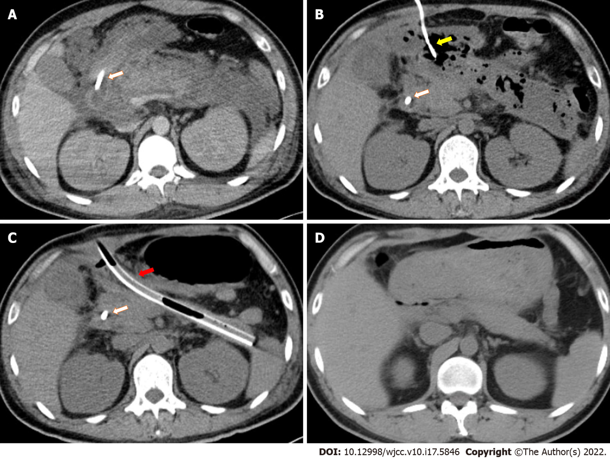
Figure 1 Pancreatic imaging changes during the course of disease.
The jejunal feeding tube is marked by a white arrow. The percutaneous drainage tube for the pancreatic head region is marked by a yellow arrow. A: Contrast-enhanced computed tomography (CT) demonstrating pancreatic edema and profound peripancreatic exudation after severe acute pancreatitis (SAP) onset; B: CT demonstrating peripancreatic infected necrosis 2 mo after SAP onset; C: CT after nephroscopy-assisted debridement of peripancreatic necrosis 3 mo after SAP onset. One of the thicker drainage tubes is marked by a red arrow; D: CT demonstrating recovery 10 mo after SAP onset.
- Citation: Wang QP, Chen YJ, Sun MX, Dai JY, Cao J, Xu Q, Zhang GN, Zhang SY. Spontaneous gallbladder perforation and colon fistula in hypertriglyceridemia-related severe acute pancreatitis: A case report. World J Clin Cases 2022; 10(17): 5846-5853
- URL: https://www.wjgnet.com/2307-8960/full/v10/i17/5846.htm
- DOI: https://dx.doi.org/10.12998/wjcc.v10.i17.5846


