Copyright
©The Author(s) 2022.
World J Clin Cases. Jun 16, 2022; 10(17): 5810-5815
Published online Jun 16, 2022. doi: 10.12998/wjcc.v10.i17.5810
Published online Jun 16, 2022. doi: 10.12998/wjcc.v10.i17.5810
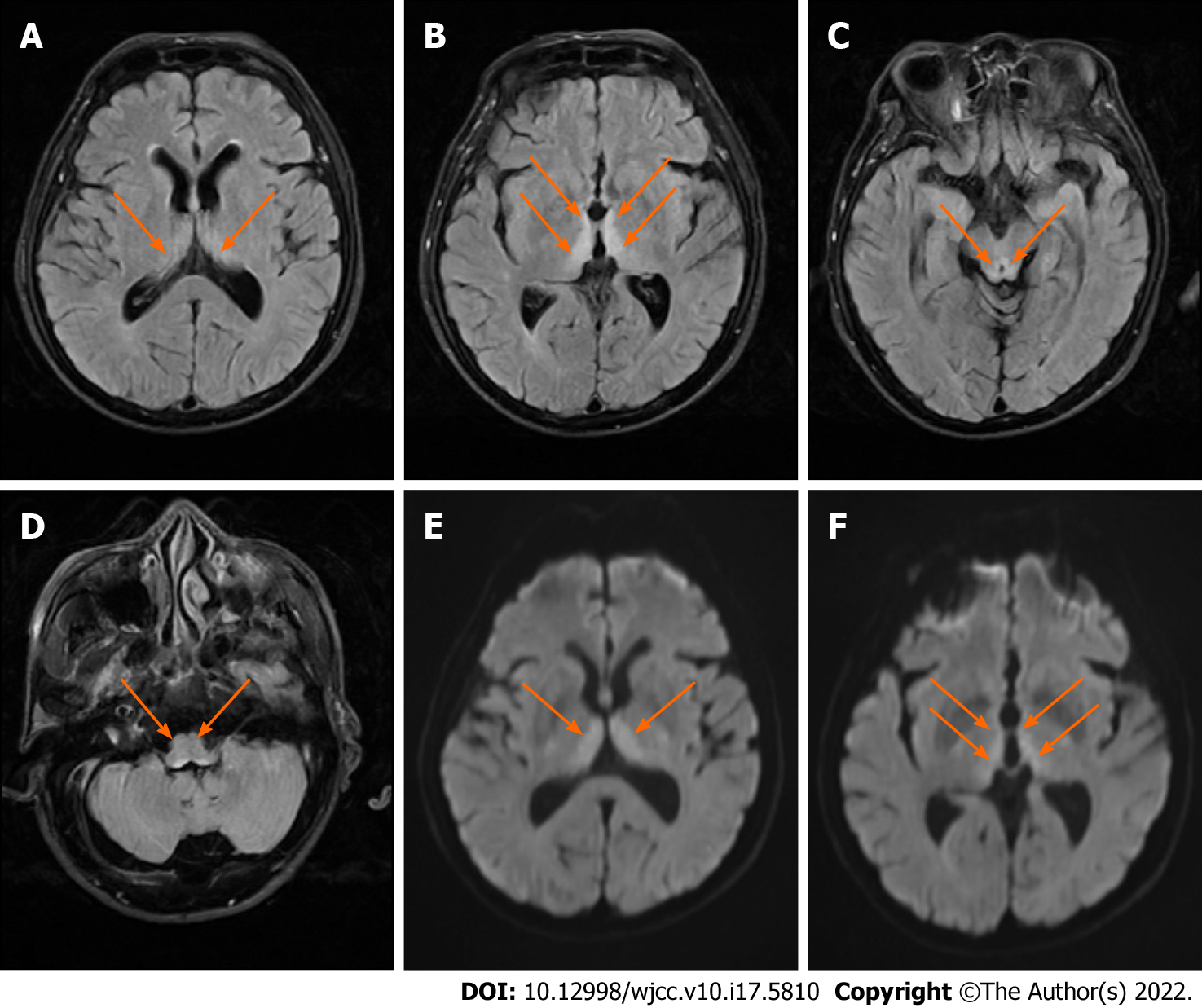
Figure 1 Patient's brain magnetic resonance imaging.
A-D: Axial fluid-attenuated inversion recovery images showing symmetrical high signals in bilateral dorsal thalami (A and B), around the third ventricle (B), cerebral aqueduct (C), and the fourth ventricle (D); E and F: Axial diffusion-weighted images showing symmetrical high signals in bilateral dorsal thalami (E and F) and the periventricular region of the third ventricle (F).
- Citation: Zhang Y, Wang L, Jiang J, Chen WY. Non-alcoholic Wernicke encephalopathy in an esophageal cancer patient receiving radiotherapy: A case report. World J Clin Cases 2022; 10(17): 5810-5815
- URL: https://www.wjgnet.com/2307-8960/full/v10/i17/5810.htm
- DOI: https://dx.doi.org/10.12998/wjcc.v10.i17.5810


