Copyright
©The Author(s) 2022.
World J Clin Cases. Jun 16, 2022; 10(17): 5798-5804
Published online Jun 16, 2022. doi: 10.12998/wjcc.v10.i17.5798
Published online Jun 16, 2022. doi: 10.12998/wjcc.v10.i17.5798
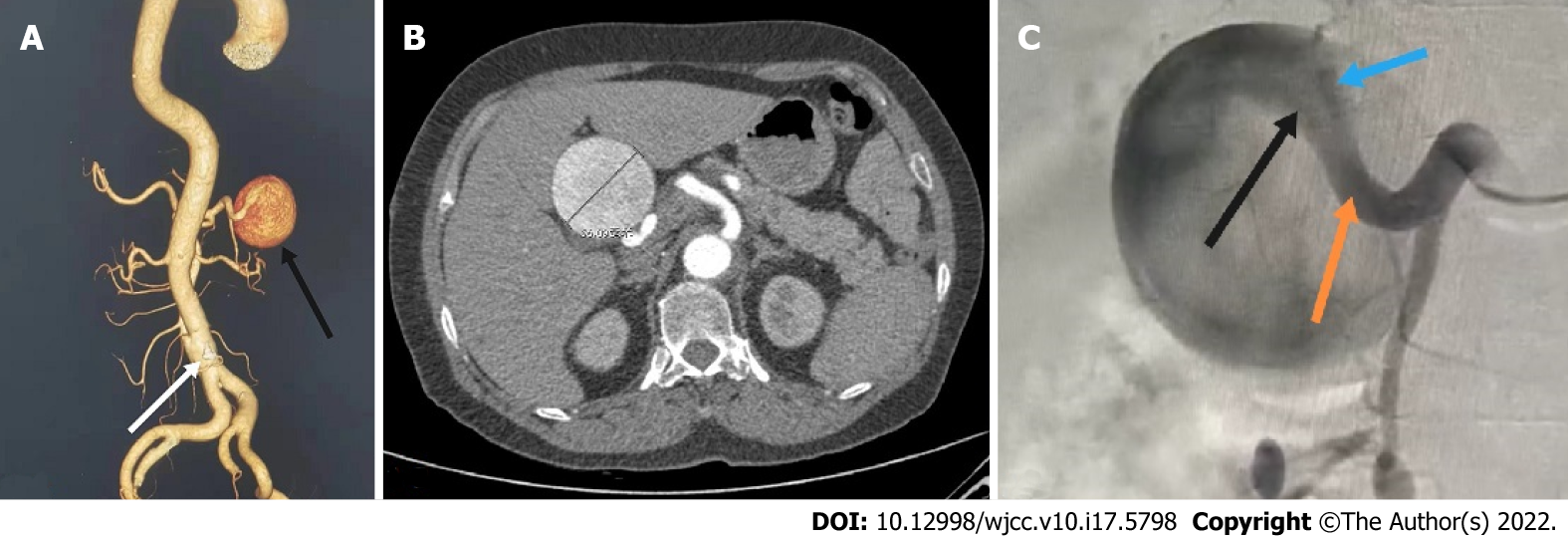
Figure 1 Computed tomography scan.
A: Abdominal computed tomography (CT) three-dimensional reconstruction showed a proper hepatic artery aneurysm (black arrow) and abdominal aortic dissection (white arrow); B: The patient's abdominal CT scan showed a huge proper hepatic artery aneurysm with a maximum diameter of approximately 56 mm; C: Abdominal aortography showed a huge proper hepatic aneurysm: A bit twisted, no collaterals, originated from the proper hepatic artery (orange arrow) and involving the bifurcation of the left (black arrow) and right hepatic arteries (blue arrow).
- Citation: Wen X, Yao ZY, Zhang Q, Wei W, Chen XY, Huang B. Surgical repair of an emergent giant hepatic aneurysm with an abdominal aortic dissection: A case report. World J Clin Cases 2022; 10(17): 5798-5804
- URL: https://www.wjgnet.com/2307-8960/full/v10/i17/5798.htm
- DOI: https://dx.doi.org/10.12998/wjcc.v10.i17.5798


