Copyright
©The Author(s) 2022.
World J Clin Cases. Jun 16, 2022; 10(17): 5770-5775
Published online Jun 16, 2022. doi: 10.12998/wjcc.v10.i17.5770
Published online Jun 16, 2022. doi: 10.12998/wjcc.v10.i17.5770
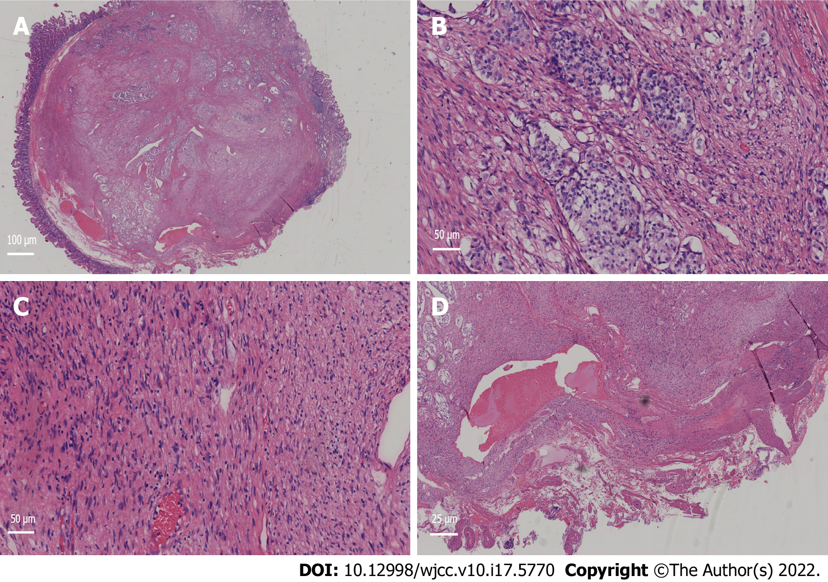
Figure 2 Pathological manifestation of the tumour under a light microscope.
A: Pathological tissue (Magnification: 100 ×). The lower right corner is the nesting tissue of the schwannoma, and the rest is the vesicle-like tissue of the neuroendocrine tumour; B: Neuroendocrine tumour tissue (Magnification: 200 ×); C: Schwannoma tissue (Magnification: 200 ×); D: The vertical incisal margin was negative, and there was no lymphatic vascular invasion (Magnification: 400 ×).
- Citation: Zhang L, Zhang C, Feng SY, Ma PP, Zhang S, Wang QQ. Neuroendocrine tumour of the descending part of the duodenum complicated with schwannoma: A case report. World J Clin Cases 2022; 10(17): 5770-5775
- URL: https://www.wjgnet.com/2307-8960/full/v10/i17/5770.htm
- DOI: https://dx.doi.org/10.12998/wjcc.v10.i17.5770


