Copyright
©The Author(s) 2022.
World J Clin Cases. Jun 16, 2022; 10(17): 5680-5689
Published online Jun 16, 2022. doi: 10.12998/wjcc.v10.i17.5680
Published online Jun 16, 2022. doi: 10.12998/wjcc.v10.i17.5680
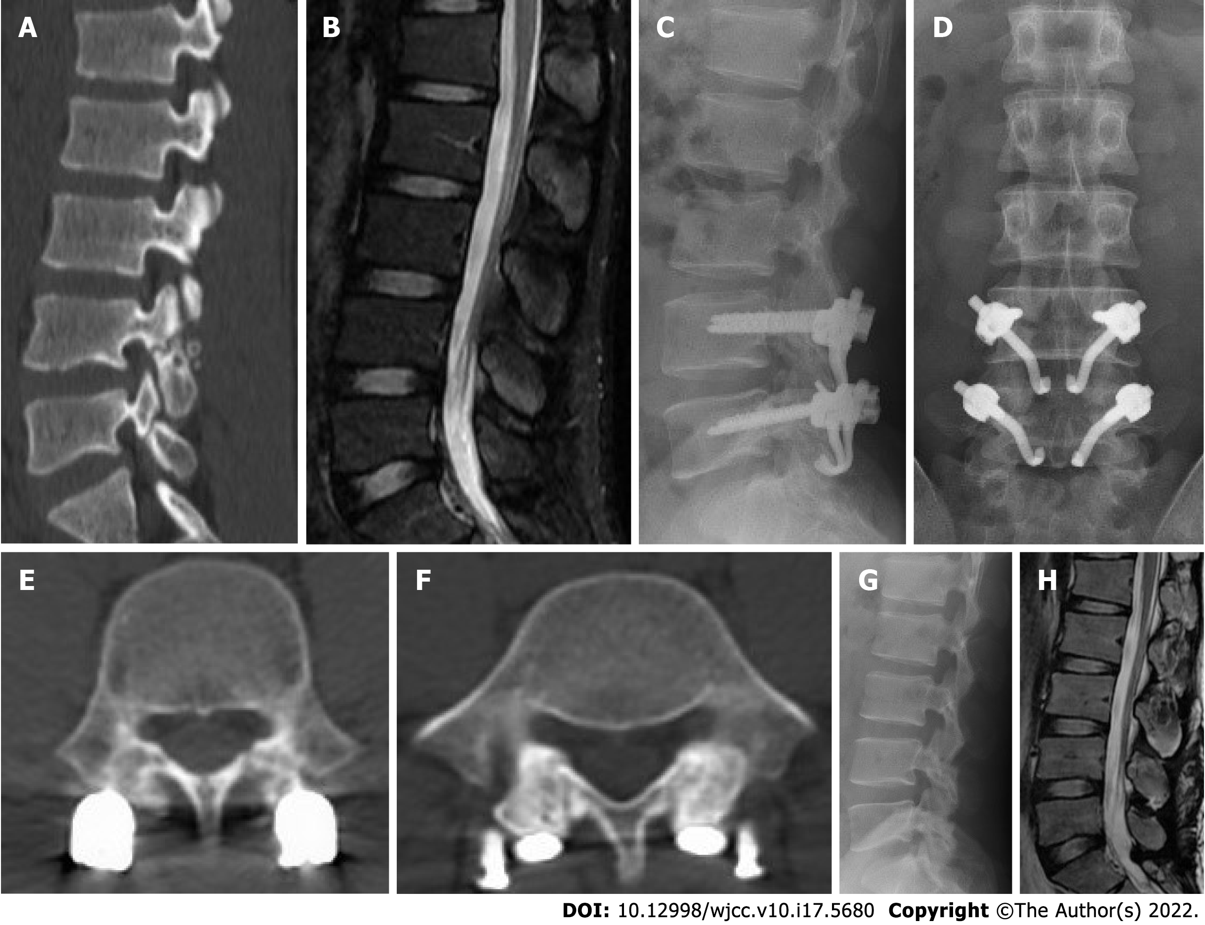
Figure 4 Typical case.
A 21-yr-old male patient had recurrent low back pain for more than 2 yr. A: Two-dimensional computed tomography scan showed lumbar spondylolysis at bilateral L4 and L5 levels; B: Lumbar magnetic resonance imaging showed normal signals of all lumbar intervertebral discs; C: Lateral radiograph after lumbar surgery; D: Anteroposterior radiograph of lumbar spine after operation; E: Computed tomography scan of the lumbar spine at 6 mo after lumbar operation showed the healing of the bone graft in the L4 isthmus; F: Computed tomography scan of the lumbar spine at 6 mo after lumbar operation showed the healing of the bone graft in the L5 isthmus; G: 12 mo after the operation the lateral radiograph showed that the internal fixation had been removed; H: 12 mo after the operation, magnetic resonance imaging showed that the signals of all lumbar intervertebral discs were normal.
- Citation: Li DM, Li YC, Jiang W, Peng BG. Application of a new anatomic hook-rod-pedicle screw system in young patients with lumbar spondylolysis: A pilot study. World J Clin Cases 2022; 10(17): 5680-5689
- URL: https://www.wjgnet.com/2307-8960/full/v10/i17/5680.htm
- DOI: https://dx.doi.org/10.12998/wjcc.v10.i17.5680


