Copyright
©The Author(s) 2022.
World J Clin Cases. Jun 6, 2022; 10(16): 5502-5509
Published online Jun 6, 2022. doi: 10.12998/wjcc.v10.i16.5502
Published online Jun 6, 2022. doi: 10.12998/wjcc.v10.i16.5502
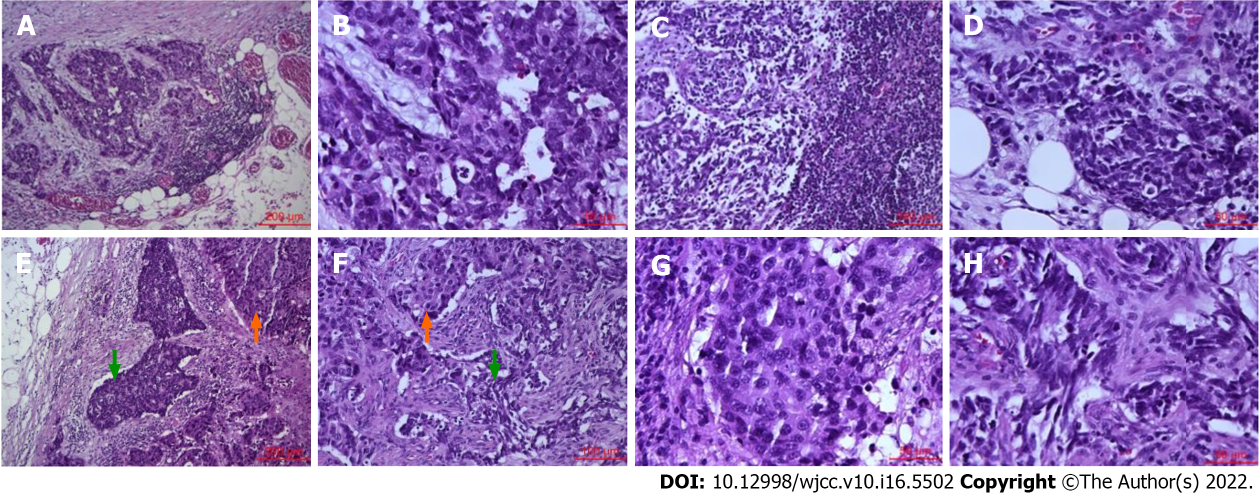
Figure 4 Histological findings of metastatic lymph nodes.
A and B: Metastatic lymph node with large cell neuroendocrine carcinoma [A, hematoxylin-eosin (HE), × 100; B, HE, × 400]; C and D: Metastatic lymph node with small cell neuroendocrine carcinoma (C, HE, × 200; D, HE, × 400); E and F: Metastatic lymph node with mixed large and small cell neuroendocrine carcinoma. The orange arrow points to large cell neuroendocrine carcinoma, and the green arrow points to small cell neuroendocrine carcinoma (E, HE, × 100; F, HE, × 200); G: Large cell neuroendocrine carcinoma components of mixed large and small cell neuroendocrine carcinoma lymph nodes (HE, × 400); H: Small cell neuroendocrine carcinoma components of mixed large and small cell neuroendocrine carcinoma lymph nodes (HE, × 400).
- Citation: Li ZF, Lu HZ, Chen YT, Bai XF, Wang TB, Fei H, Zhao DB. Mixed large and small cell neuroendocrine carcinoma of the stomach: A case report and review of literature. World J Clin Cases 2022; 10(16): 5502-5509
- URL: https://www.wjgnet.com/2307-8960/full/v10/i16/5502.htm
- DOI: https://dx.doi.org/10.12998/wjcc.v10.i16.5502


