Copyright
©The Author(s) 2022.
World J Clin Cases. Jun 6, 2022; 10(16): 5470-5478
Published online Jun 6, 2022. doi: 10.12998/wjcc.v10.i16.5470
Published online Jun 6, 2022. doi: 10.12998/wjcc.v10.i16.5470
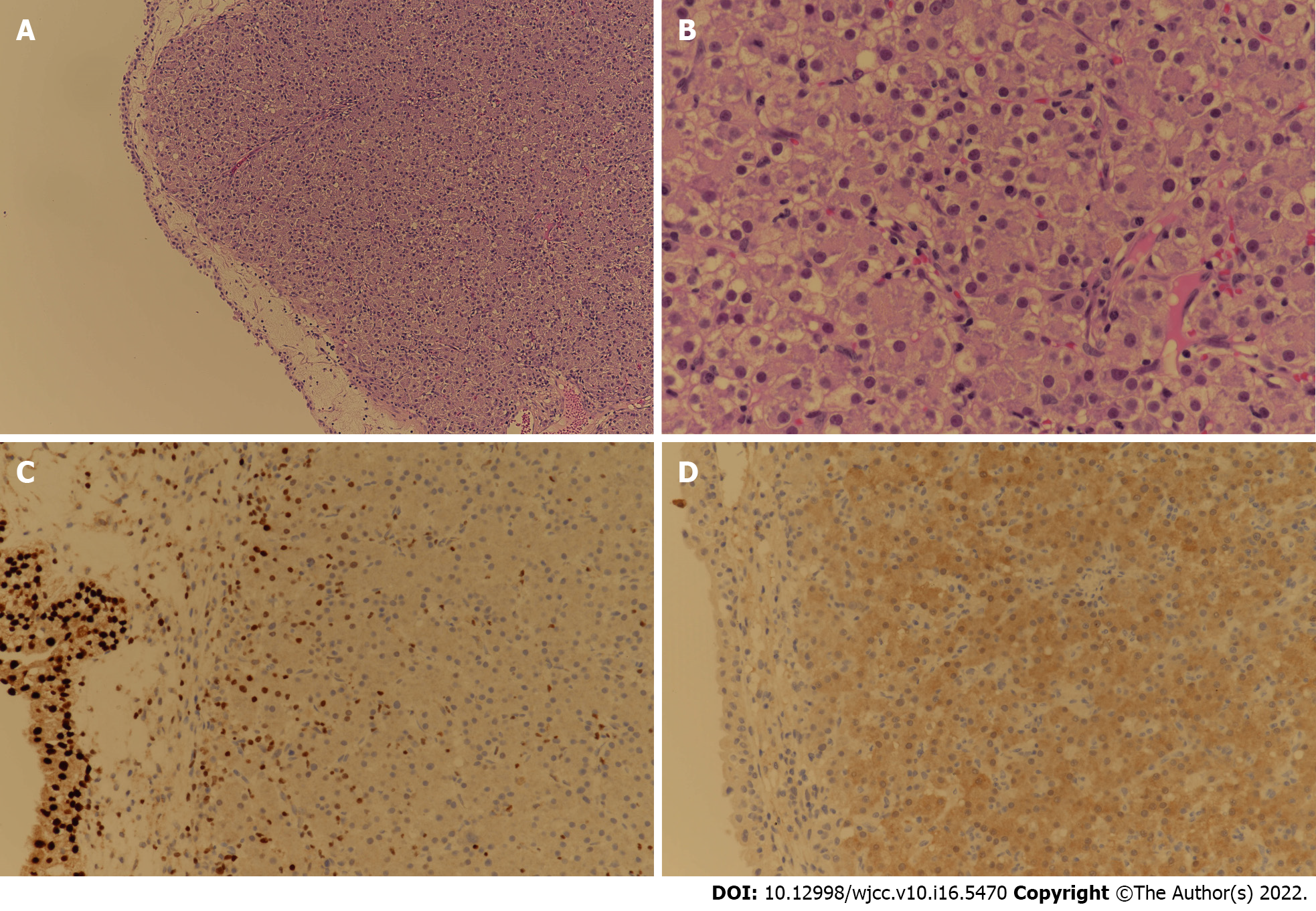
Figure 5 Tissue and immunohistochemical staining results of the bladder tumor tissue resected by transurethral resection of bladder tumor.
A: Tumor cells show solid sheet architecture in HE staining (× 100); B: Tumor cells are polygonal with nuclear atypia in HE staining (× 400); C: GATA binding protein 3 staining (× 200) is negative for tumor cells; D: Tumor cells are positive in Arginase-1 staining (× 200), which is compatible with hepatocellular carcinoma.
- Citation: Kim Y, Kim YS, Yoo JJ, Kim SG, Chin S, Moon A. Rare case of hepatocellular carcinoma metastasis to urinary bladder: A case report . World J Clin Cases 2022; 10(16): 5470-5478
- URL: https://www.wjgnet.com/2307-8960/full/v10/i16/5470.htm
- DOI: https://dx.doi.org/10.12998/wjcc.v10.i16.5470


