Copyright
©The Author(s) 2022.
World J Clin Cases. Jun 6, 2022; 10(16): 5420-5427
Published online Jun 6, 2022. doi: 10.12998/wjcc.v10.i16.5420
Published online Jun 6, 2022. doi: 10.12998/wjcc.v10.i16.5420
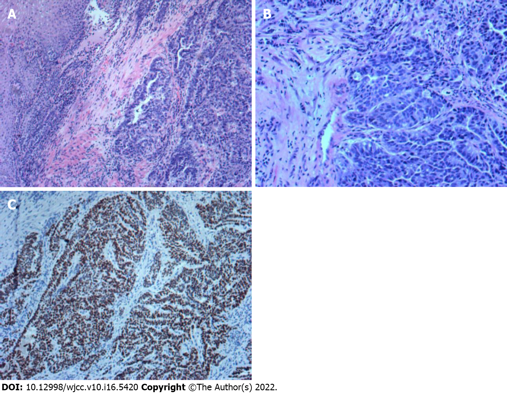
Figure 2 Histological analysis of the patient’s tumour tissue.
A: Hematoxylin and eosin staining: tumor cells are arranged in a trabecular patten, with a glandular and hepatoid component (magnification, ×100); B: Hematoxylin and eosin staining: most of them are hepatocyte like differentiation area, and a few are adenocarcinoma area (magnification, ×200); C: Immunohistochemical staining: cells are positively stained for SALL4 (magnification, ×100).
- Citation: Liu M, Luo C, Xie ZZ, Li X. Treatment of gastric hepatoid adenocarcinoma with pembrolizumab and bevacizumab combination chemotherapy: A case report. World J Clin Cases 2022; 10(16): 5420-5427
- URL: https://www.wjgnet.com/2307-8960/full/v10/i16/5420.htm
- DOI: https://dx.doi.org/10.12998/wjcc.v10.i16.5420


