Copyright
©The Author(s) 2022.
World J Clin Cases. Jun 6, 2022; 10(16): 5406-5413
Published online Jun 6, 2022. doi: 10.12998/wjcc.v10.i16.5406
Published online Jun 6, 2022. doi: 10.12998/wjcc.v10.i16.5406
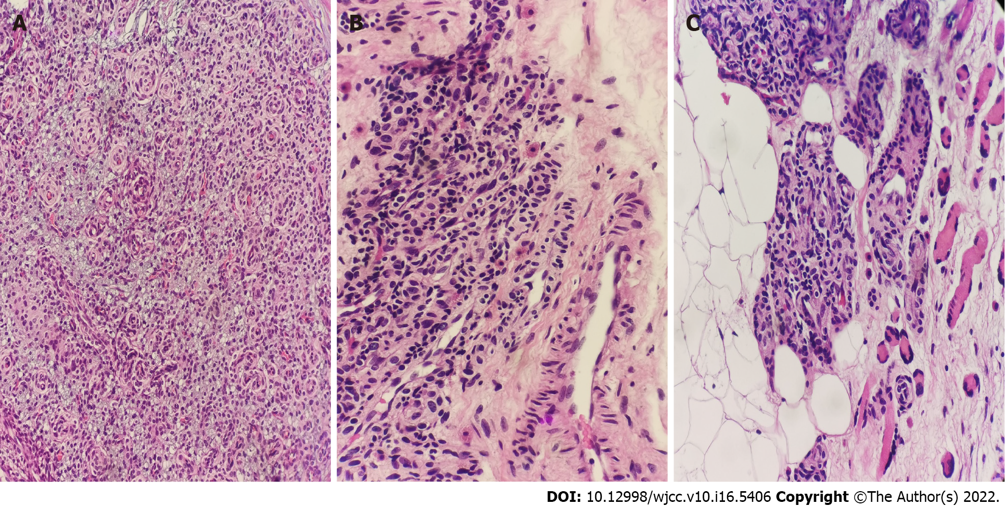
Figure 2 Images under the microscope.
A: Diffuse proliferation of small vessels with oval cell proliferation around, mild cell morphology, abundant cytoplasm, close relationship with blood vessels, no obvious atypia, and clear mitosis (hematoxylin and eosin 10 ×); B: Oval cells beside small vessels can be seen in the surrounding tissues, which is consistent with the shape of tumor cells. Mast cells are scattered in the stroma. (Hematoxylin and eosin 20 ×); C: The tumor infiltrates the surrounding adipose connective tissue. (Hematoxylin and eosin 20 ×).
- Citation: Wu RC, Gao YH, Sun WW, Zhang XY, Zhang SP. Glomangiomatosis - immunohistochemical study: A case report. World J Clin Cases 2022; 10(16): 5406-5413
- URL: https://www.wjgnet.com/2307-8960/full/v10/i16/5406.htm
- DOI: https://dx.doi.org/10.12998/wjcc.v10.i16.5406


