Copyright
©The Author(s) 2022.
World J Clin Cases. Jun 6, 2022; 10(16): 5343-5351
Published online Jun 6, 2022. doi: 10.12998/wjcc.v10.i16.5343
Published online Jun 6, 2022. doi: 10.12998/wjcc.v10.i16.5343
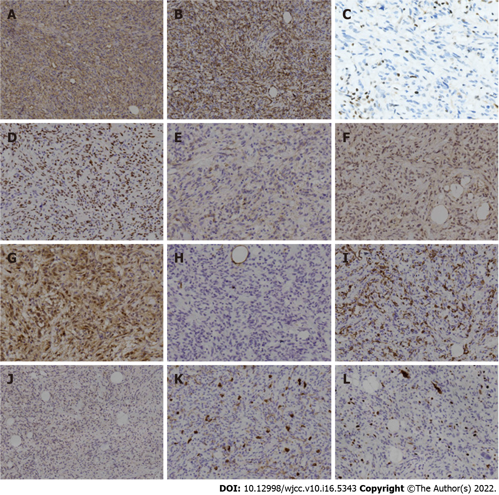
Figure 4 Immunohistochemical findings in the tumor.
A: Cells showed strongly positive staining for cluster of differentiation (CD) 34; B: Cells showed strongly positive staining for desmin; C: Lost expression of retinoblastoma 1 protein, with positive internal-control staining for lymphocytes; D: Staining was positive for estrogen receptor; E: Staining was positive for epithelial-membrane antigen; F: Staining was positive for murine double minute 2; G: Staining was positive for cyclin-dependent kinase 4; H: Staining was negative for S100; I: Staining was negative for smooth muscle actin; J: Staining was negative for signal transducer and activator of transcription 6; K: Staining was negative for CD117. Note that positive staining for CD117 appeared in the mast cells of the internal control; L: Ki-67 index was about 5%.
- Citation: Zeng YF, Dai YZ, Chen M. Mammary-type myofibroblastoma with infarction and atypical mitosis-a potential diagnostic pitfall: A case report. World J Clin Cases 2022; 10(16): 5343-5351
- URL: https://www.wjgnet.com/2307-8960/full/v10/i16/5343.htm
- DOI: https://dx.doi.org/10.12998/wjcc.v10.i16.5343


