Copyright
©The Author(s) 2022.
World J Clin Cases. Jun 6, 2022; 10(16): 5343-5351
Published online Jun 6, 2022. doi: 10.12998/wjcc.v10.i16.5343
Published online Jun 6, 2022. doi: 10.12998/wjcc.v10.i16.5343
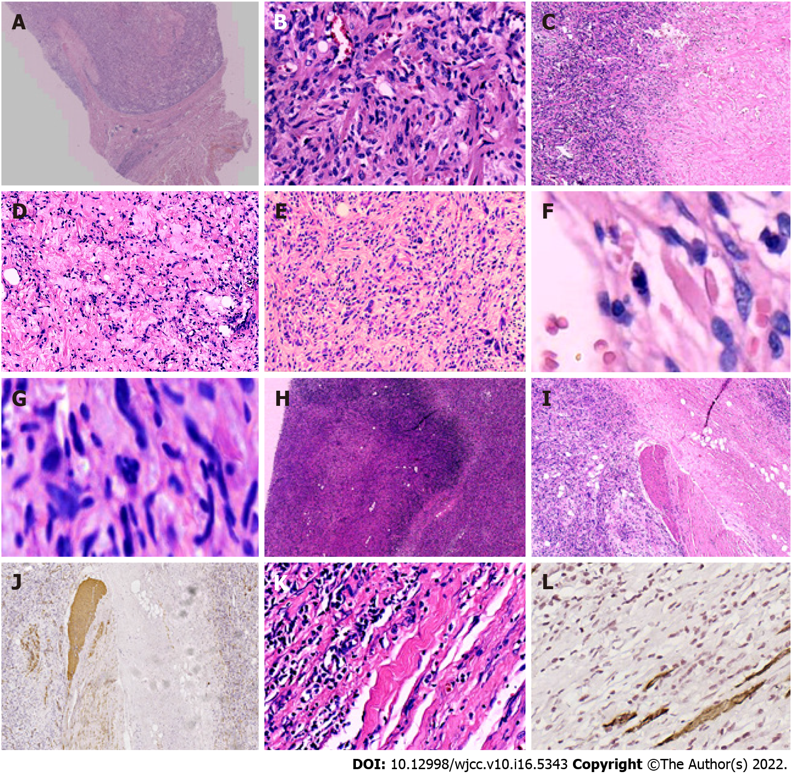
Figure 3 Histopathological features of the tumor.
A: The mass had a definite capsule; B: It was composed of short spindle to oval-shaped cells, and admixed with varying thick bundles of collagen and variable numbers of adipocytes. The cells had eosinophilic cytoplasm with indistinct cell borders and elongated nuclei with fine chromatin; C: Hyaline degeneration was visible within the tumor; D: Mucoid degeneration was visible within the tumor; E: Bizarre cells and atypic cells were unequally distributed throughout the tumor; F: Mitosis was found in the tumor cells; G: Atypical mitosis was found in the tumor cells; H: Infarction and nuclear debris were also seen inside the tumor; I and J: Smooth muscles had been infiltrated by the tumor cells; K and L: Skeletal muscles had been infiltrated by the tumor cells. Images I and K show local magnification of the black and blue boxes, respectively, in Figure A. Image J shows the infiltrated smooth muscle, which was positive for smooth muscle actin. Image L displays the invaded skeletal muscle, which was positive for desmin on immunohistochemical staining.
- Citation: Zeng YF, Dai YZ, Chen M. Mammary-type myofibroblastoma with infarction and atypical mitosis-a potential diagnostic pitfall: A case report. World J Clin Cases 2022; 10(16): 5343-5351
- URL: https://www.wjgnet.com/2307-8960/full/v10/i16/5343.htm
- DOI: https://dx.doi.org/10.12998/wjcc.v10.i16.5343


