Copyright
©The Author(s) 2022.
World J Clin Cases. Jun 6, 2022; 10(16): 5343-5351
Published online Jun 6, 2022. doi: 10.12998/wjcc.v10.i16.5343
Published online Jun 6, 2022. doi: 10.12998/wjcc.v10.i16.5343
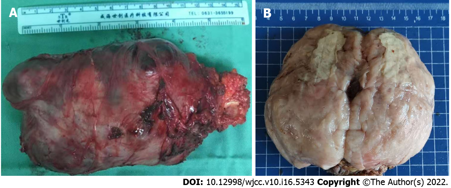
Figure 2 General observation of the mass.
A: Postoperative gross image demonstrates that the mass had an almost intact capsule with the maximum diameter of about 13 cm in size; B: General sectional view displays that the mass was solid, firm-to-elastic, and yellow-pink appearance, with focal cystic degeneration and necrosis.
- Citation: Zeng YF, Dai YZ, Chen M. Mammary-type myofibroblastoma with infarction and atypical mitosis-a potential diagnostic pitfall: A case report. World J Clin Cases 2022; 10(16): 5343-5351
- URL: https://www.wjgnet.com/2307-8960/full/v10/i16/5343.htm
- DOI: https://dx.doi.org/10.12998/wjcc.v10.i16.5343


