Copyright
©The Author(s) 2022.
World J Clin Cases. Jun 6, 2022; 10(16): 5185-5195
Published online Jun 6, 2022. doi: 10.12998/wjcc.v10.i16.5185
Published online Jun 6, 2022. doi: 10.12998/wjcc.v10.i16.5185
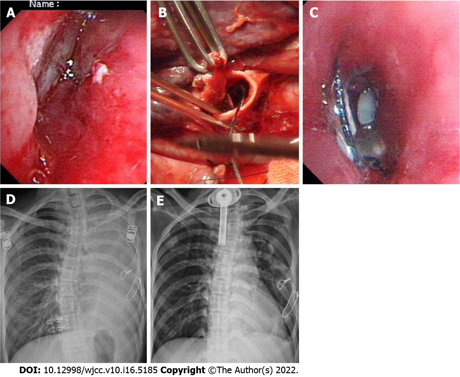
Figure 5 Case 9.
A: Fiberoptic bronchoscopy (FB) showed deformed and collapsed left main bronchus the day after injury; B: Intraoperative image showing transected bronchus, debridement, and end-to-end anastomosis; C: FB showed lumen stenosis with sutures across 3 wk after surgery; D: FB failed to relieve the stenosis and repeated atelectasis; E: Reoperation was performed for resection of stenosis and anastomosis 3 years later, and postoperative computed tomography shows distended left lung.
- Citation: Gao JM, Li H, Du DY, Yang J, Kong LW, Wang JB, He P, Wei GB. Management and outcome of bronchial trauma due to blunt versus penetrating injuries. World J Clin Cases 2022; 10(16): 5185-5195
- URL: https://www.wjgnet.com/2307-8960/full/v10/i16/5185.htm
- DOI: https://dx.doi.org/10.12998/wjcc.v10.i16.5185


