Copyright
©The Author(s) 2022.
World J Clin Cases. Jun 6, 2022; 10(16): 5185-5195
Published online Jun 6, 2022. doi: 10.12998/wjcc.v10.i16.5185
Published online Jun 6, 2022. doi: 10.12998/wjcc.v10.i16.5185
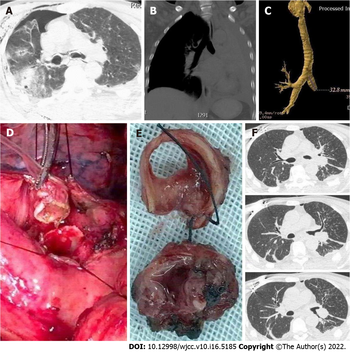
Figure 4 Case 8.
A: Computed tomography (CT) image showing a deformed left main bronchus, discontinuous with air around bronchi, besides the right pneumothorax and subcutaneous emphysema at admission; B and C: After 50 d, CT showed left atelectasis and the interrupted left main bronchus; D and E: Intraoperative images showing transected bronchus and scar excision followed by end-to-end anastomosis; F: Postoperative CT image showing distended left lung, and unblocked main bronchus and lobar bronchi.
- Citation: Gao JM, Li H, Du DY, Yang J, Kong LW, Wang JB, He P, Wei GB. Management and outcome of bronchial trauma due to blunt versus penetrating injuries. World J Clin Cases 2022; 10(16): 5185-5195
- URL: https://www.wjgnet.com/2307-8960/full/v10/i16/5185.htm
- DOI: https://dx.doi.org/10.12998/wjcc.v10.i16.5185


