Copyright
©The Author(s) 2022.
World J Clin Cases. May 26, 2022; 10(15): 5064-5071
Published online May 26, 2022. doi: 10.12998/wjcc.v10.i15.5064
Published online May 26, 2022. doi: 10.12998/wjcc.v10.i15.5064
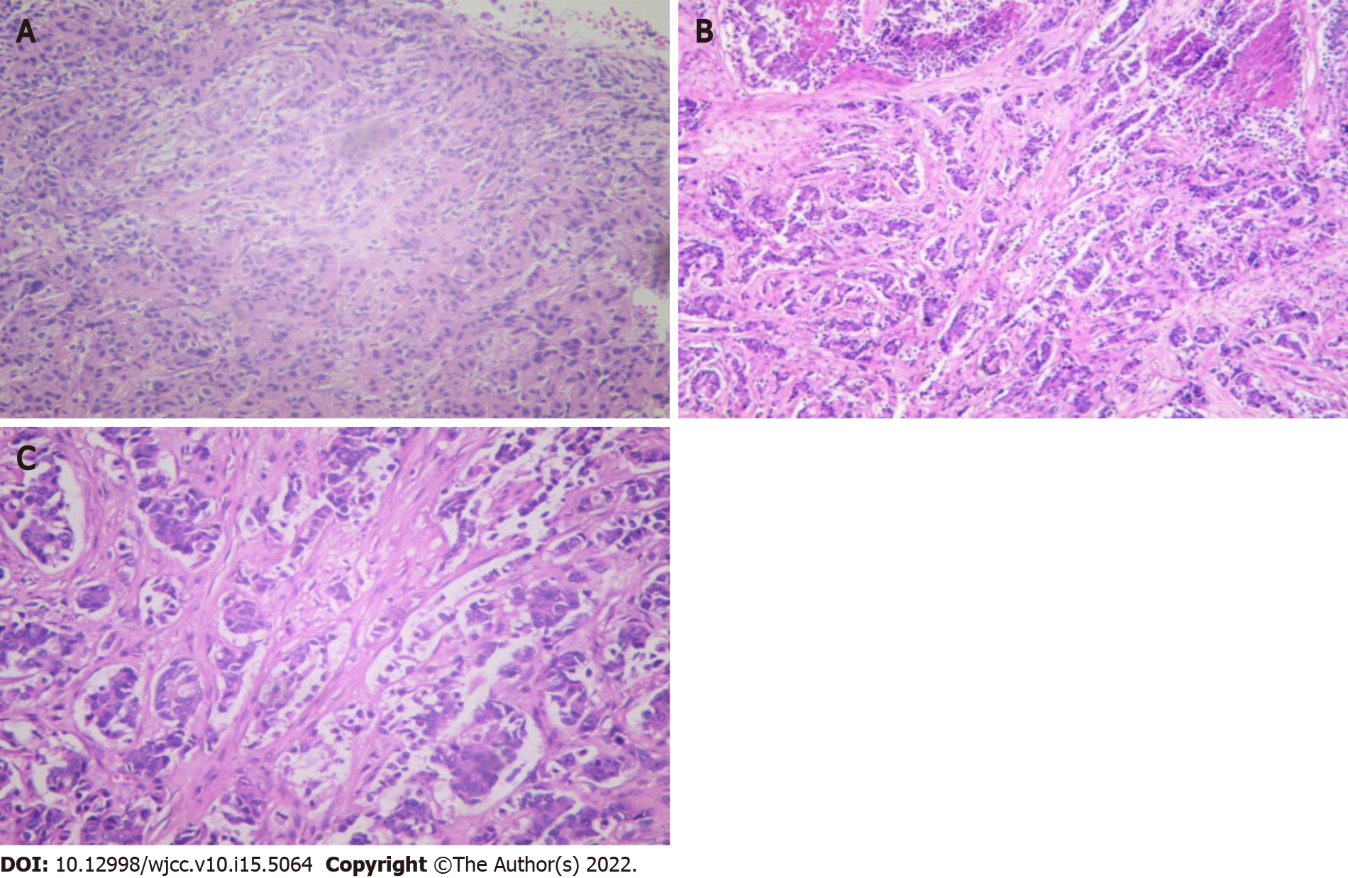
Figure 3 Core needle biopsy of both breast masses.
A: Fine needle aspiration cytology to identify tumor cells (4 × 10); B: An immunohistochemical pathological section of invasive ductal carcinoma in the left breast (4 × 4); C: An immunohistochemical pathological section of invasive ductal carcinoma in the right breast (4 × 10).
- Citation: Yang SY, Li Y, Nie JY, Yang ST, Yang XJ, Wang MH, Zhang J. Metaplastic breast cancer with chondrosarcomatous differentiation combined with concurrent bilateral breast cancer: A case report. World J Clin Cases 2022; 10(15): 5064-5071
- URL: https://www.wjgnet.com/2307-8960/full/v10/i15/5064.htm
- DOI: https://dx.doi.org/10.12998/wjcc.v10.i15.5064


