Copyright
©The Author(s) 2022.
World J Clin Cases. May 26, 2022; 10(15): 5042-5050
Published online May 26, 2022. doi: 10.12998/wjcc.v10.i15.5042
Published online May 26, 2022. doi: 10.12998/wjcc.v10.i15.5042
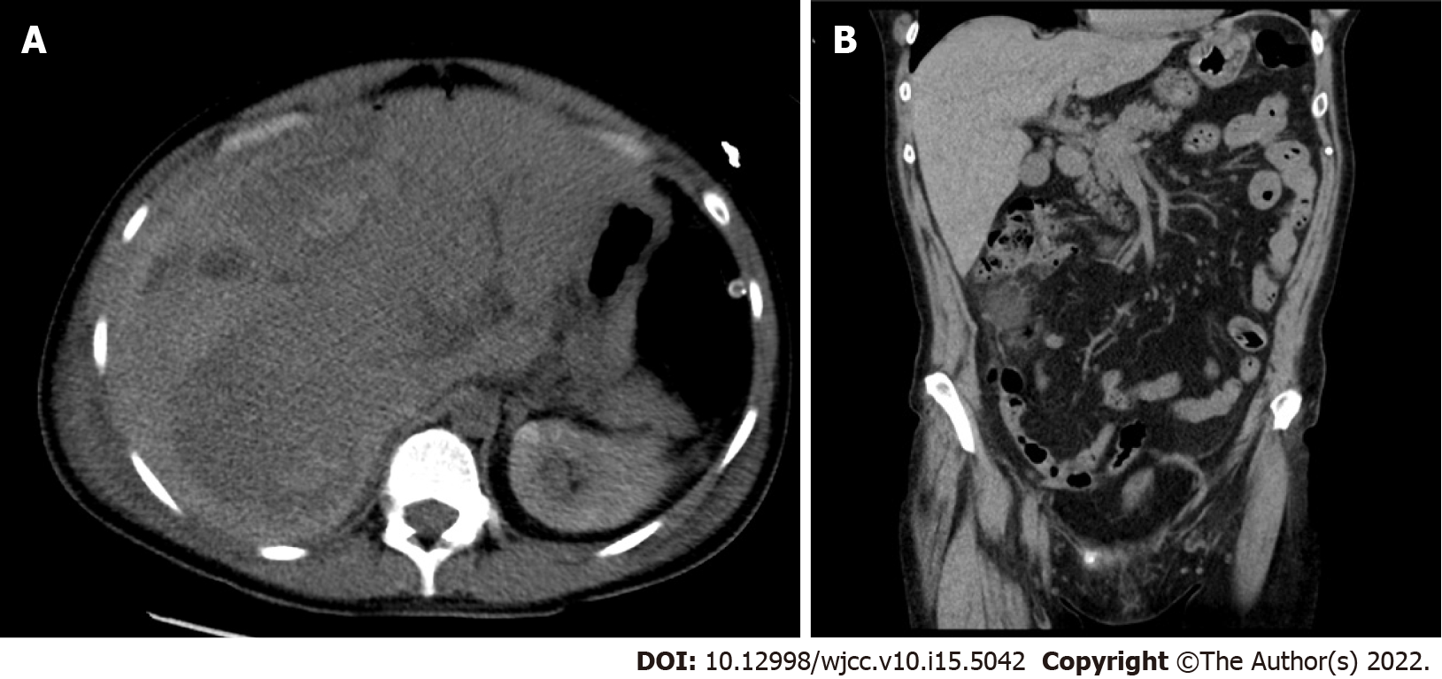
Figure 1 Abdominal computed tomography with intravenous contrast.
A: Transverse section: right hepatic lobe haematoma (158 mm × 94 mm); B: Sagittal section: Right hepatic lobe haematoma, craniocaudal size 240 mm.
- Citation: Ambrož R, Stašek M, Molnár J, Špička P, Klos D, Hambálek J, Skanderová D. Spontaneous liver rupture following SARS-CoV-2 infection in late pregnancy: A case report. World J Clin Cases 2022; 10(15): 5042-5050
- URL: https://www.wjgnet.com/2307-8960/full/v10/i15/5042.htm
- DOI: https://dx.doi.org/10.12998/wjcc.v10.i15.5042


