Copyright
©The Author(s) 2022.
World J Clin Cases. May 26, 2022; 10(15): 4971-4984
Published online May 26, 2022. doi: 10.12998/wjcc.v10.i15.4971
Published online May 26, 2022. doi: 10.12998/wjcc.v10.i15.4971
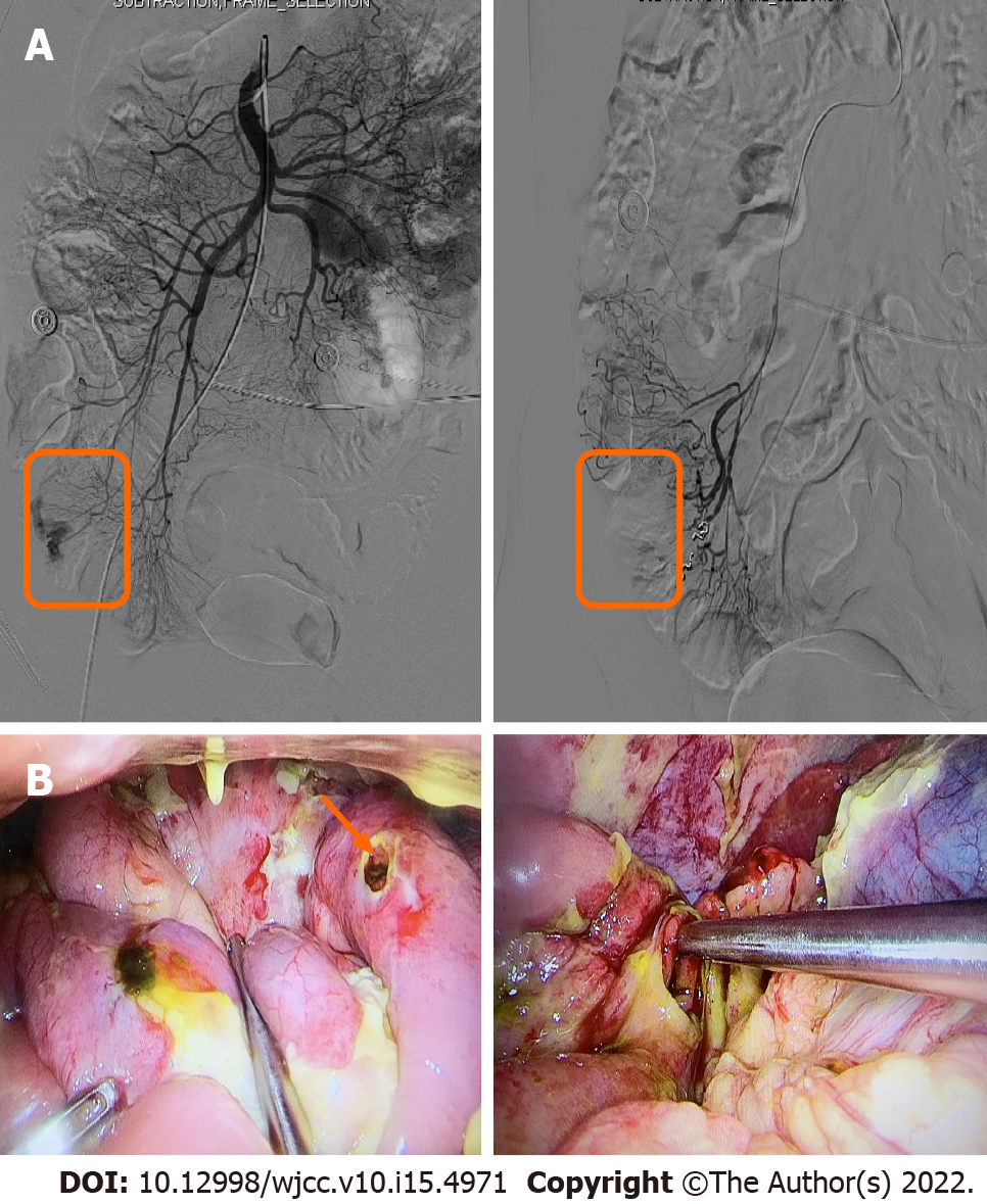
Figure 3 Superior mesenteric arteriography and perioperative images.
A: Superior mesenteric arteriography shows rupture and hemorrhage of a straight arteriole distal to the ileocolic artery; subsequently, microspring coils are used to embolize the diseased vessels, and repeated angiography shows that the hemorrhagic lesion disappears 5 minutes later (Red box); B: A large amount of yellow-green intestinal fluid in the abdominal cavity and ileum perforation are observed during the operation (Orange arrow).
- Citation: Weng CY, Ye C, Fan YH, Lv B, Zhang CL, Li M. CD8-positive indolent T-Cell lymphoproliferative disorder of the gastrointestinal tract: A case report and review of literature. World J Clin Cases 2022; 10(15): 4971-4984
- URL: https://www.wjgnet.com/2307-8960/full/v10/i15/4971.htm
- DOI: https://dx.doi.org/10.12998/wjcc.v10.i15.4971


