Copyright
©The Author(s) 2022.
World J Clin Cases. May 26, 2022; 10(15): 4942-4948
Published online May 26, 2022. doi: 10.12998/wjcc.v10.i15.4942
Published online May 26, 2022. doi: 10.12998/wjcc.v10.i15.4942
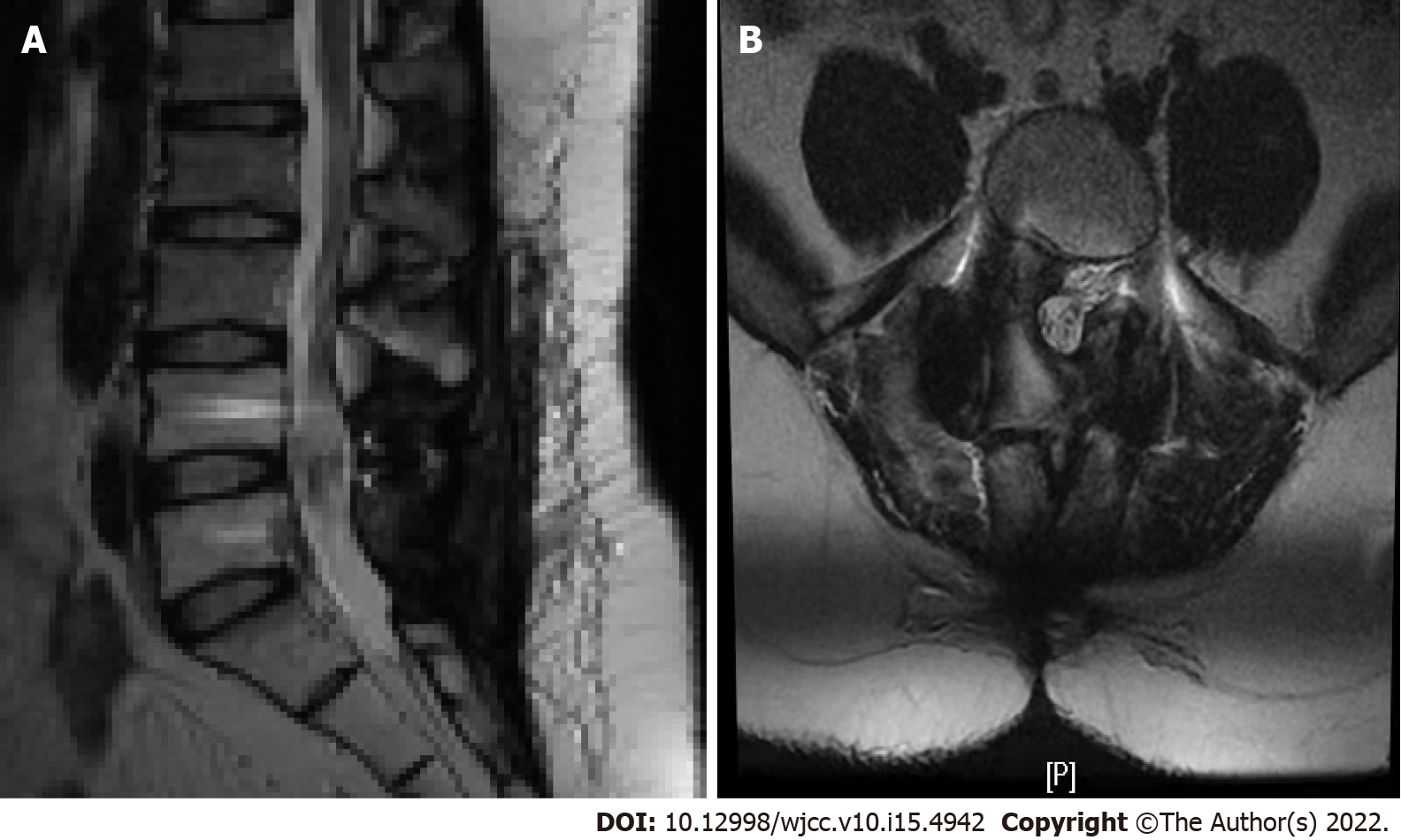
Figure 3 Magnetic resonance imaging examination after 1 year and 3 mo of follow-up.
A and B: Magnetic resonance imaging examination showed that the space occupying the spinal canal at the L4-5 level had been removed, and no obvious metabolic signs of recurrence were observed.
- Citation: Lei LH, Li F, Wu T. Primary extraskeletal Ewing’s sarcoma of the lumbar nerve root: A case report. World J Clin Cases 2022; 10(15): 4942-4948
- URL: https://www.wjgnet.com/2307-8960/full/v10/i15/4942.htm
- DOI: https://dx.doi.org/10.12998/wjcc.v10.i15.4942


