Copyright
©The Author(s) 2022.
World J Clin Cases. May 26, 2022; 10(15): 4942-4948
Published online May 26, 2022. doi: 10.12998/wjcc.v10.i15.4942
Published online May 26, 2022. doi: 10.12998/wjcc.v10.i15.4942
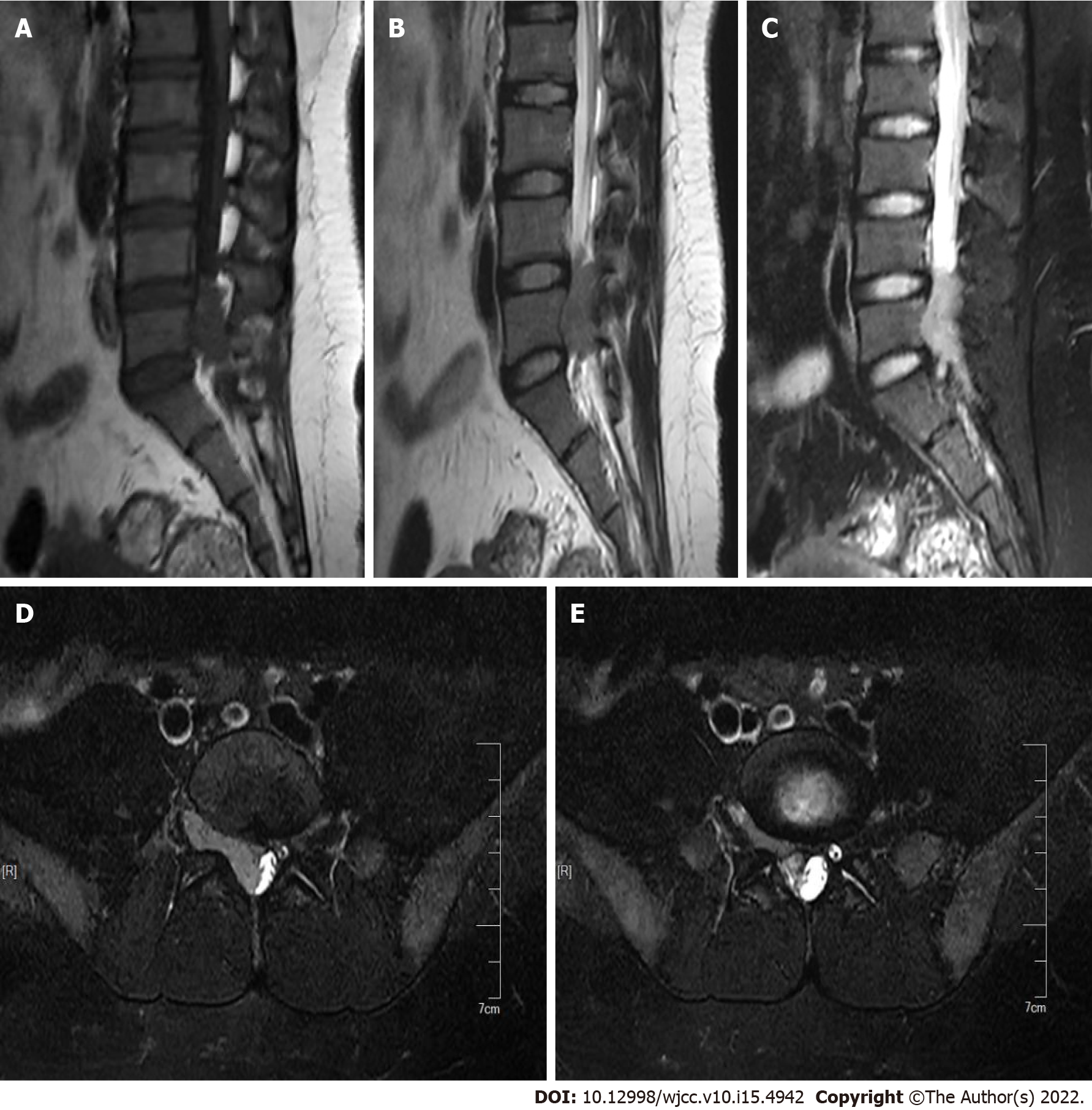
Figure 1 Preoperative magnetic resonance imaging examination.
A: Magnetic resonance imaging (MRI) shows low intensity on T1W1; B: MRI shows slightly high and slightly low intensities on T2W1, with a few small nodules of high intensity visible; C: MRI shows high intensity on STIR; D: With the mass extending into the intervertebral foramen in cross section; E: With the mass extending into the intervertebral foramen in cross section.
- Citation: Lei LH, Li F, Wu T. Primary extraskeletal Ewing’s sarcoma of the lumbar nerve root: A case report. World J Clin Cases 2022; 10(15): 4942-4948
- URL: https://www.wjgnet.com/2307-8960/full/v10/i15/4942.htm
- DOI: https://dx.doi.org/10.12998/wjcc.v10.i15.4942


