Copyright
©The Author(s) 2022.
World J Clin Cases. May 26, 2022; 10(15): 4935-4941
Published online May 26, 2022. doi: 10.12998/wjcc.v10.i15.4935
Published online May 26, 2022. doi: 10.12998/wjcc.v10.i15.4935
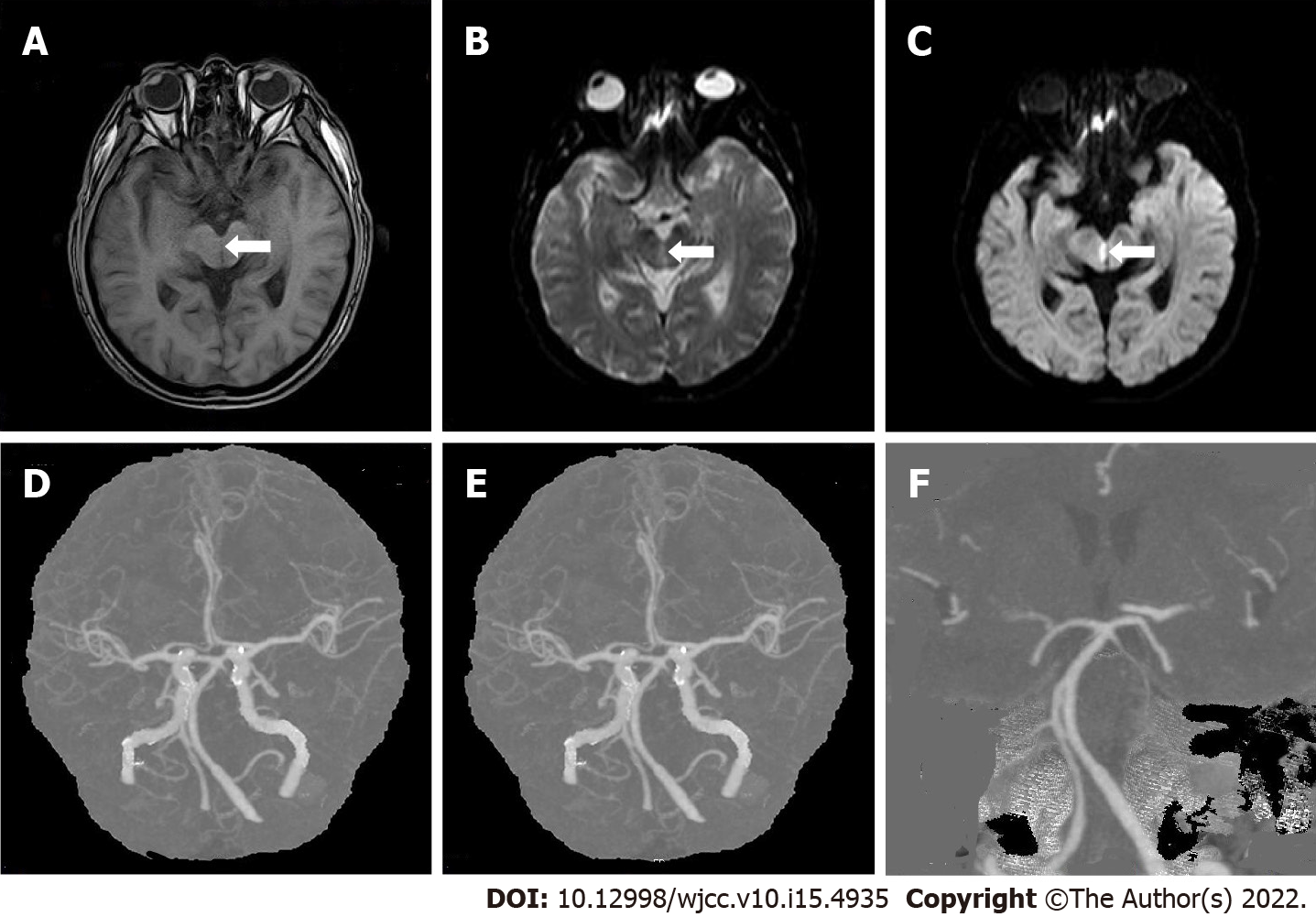
Figure 2 Patient 2.
A: Brain magnetic resonance imaging (MRI) on February 6, T1 weighted image, T1 low signal can be seen at the midbrain aqueduct; B: Brain MRI on February 6, T2 weighted image, T2 high signal can be seen at the midbrain aqueduct; C: Brain MRI on February 6, diffusion-weighted imaging, high signal can be seen in the midbrain aqueduct; D-F: Cerebrovascular computed tomography angiogram on February 13, a little mixed plaque at the bifurcation of the right common carotid artery.
- Citation: Yang YZ, Hu WX, Zhai HJ. Special type of Wernekink syndrome in midbrain infarction: Four case reports. World J Clin Cases 2022; 10(15): 4935-4941
- URL: https://www.wjgnet.com/2307-8960/full/v10/i15/4935.htm
- DOI: https://dx.doi.org/10.12998/wjcc.v10.i15.4935


