Copyright
©The Author(s) 2022.
World J Clin Cases. May 26, 2022; 10(15): 4929-4934
Published online May 26, 2022. doi: 10.12998/wjcc.v10.i15.4929
Published online May 26, 2022. doi: 10.12998/wjcc.v10.i15.4929
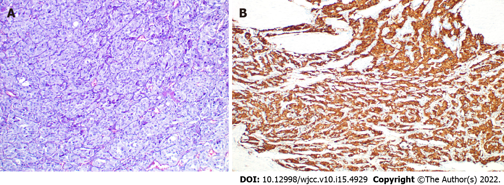
Figure 3 Pathological and immunological features of a paraganglioma of the urinary bladder.
A: Micrograph of conventional HE staining (× 200) showing chief cells in a nested growth pattern; B: The image shows positive expression of CgA in immunohistochemistry.
- Citation: Chen J, Yang HF. Nonfunctional bladder paraganglioma misdiagnosed as hemangioma: A case report. World J Clin Cases 2022; 10(15): 4929-4934
- URL: https://www.wjgnet.com/2307-8960/full/v10/i15/4929.htm
- DOI: https://dx.doi.org/10.12998/wjcc.v10.i15.4929


