Copyright
©The Author(s) 2022.
World J Clin Cases. May 26, 2022; 10(15): 4929-4934
Published online May 26, 2022. doi: 10.12998/wjcc.v10.i15.4929
Published online May 26, 2022. doi: 10.12998/wjcc.v10.i15.4929
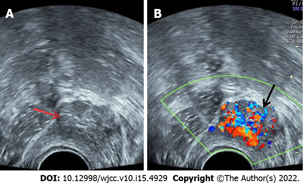
Figure 1 Transvaginal ultrasound showed a 2.
5 cm × 2.1 cm medium-echo mass protruding from the right anterior wall of the bladder (A, indicated by the red arrow); a regular shape, a clear boundary, a wide base, and rich strip blood flow signals were detected (B, indicated by the black arrow).
- Citation: Chen J, Yang HF. Nonfunctional bladder paraganglioma misdiagnosed as hemangioma: A case report. World J Clin Cases 2022; 10(15): 4929-4934
- URL: https://www.wjgnet.com/2307-8960/full/v10/i15/4929.htm
- DOI: https://dx.doi.org/10.12998/wjcc.v10.i15.4929


