Copyright
©The Author(s) 2022.
World J Clin Cases. May 26, 2022; 10(15): 4895-4903
Published online May 26, 2022. doi: 10.12998/wjcc.v10.i15.4895
Published online May 26, 2022. doi: 10.12998/wjcc.v10.i15.4895
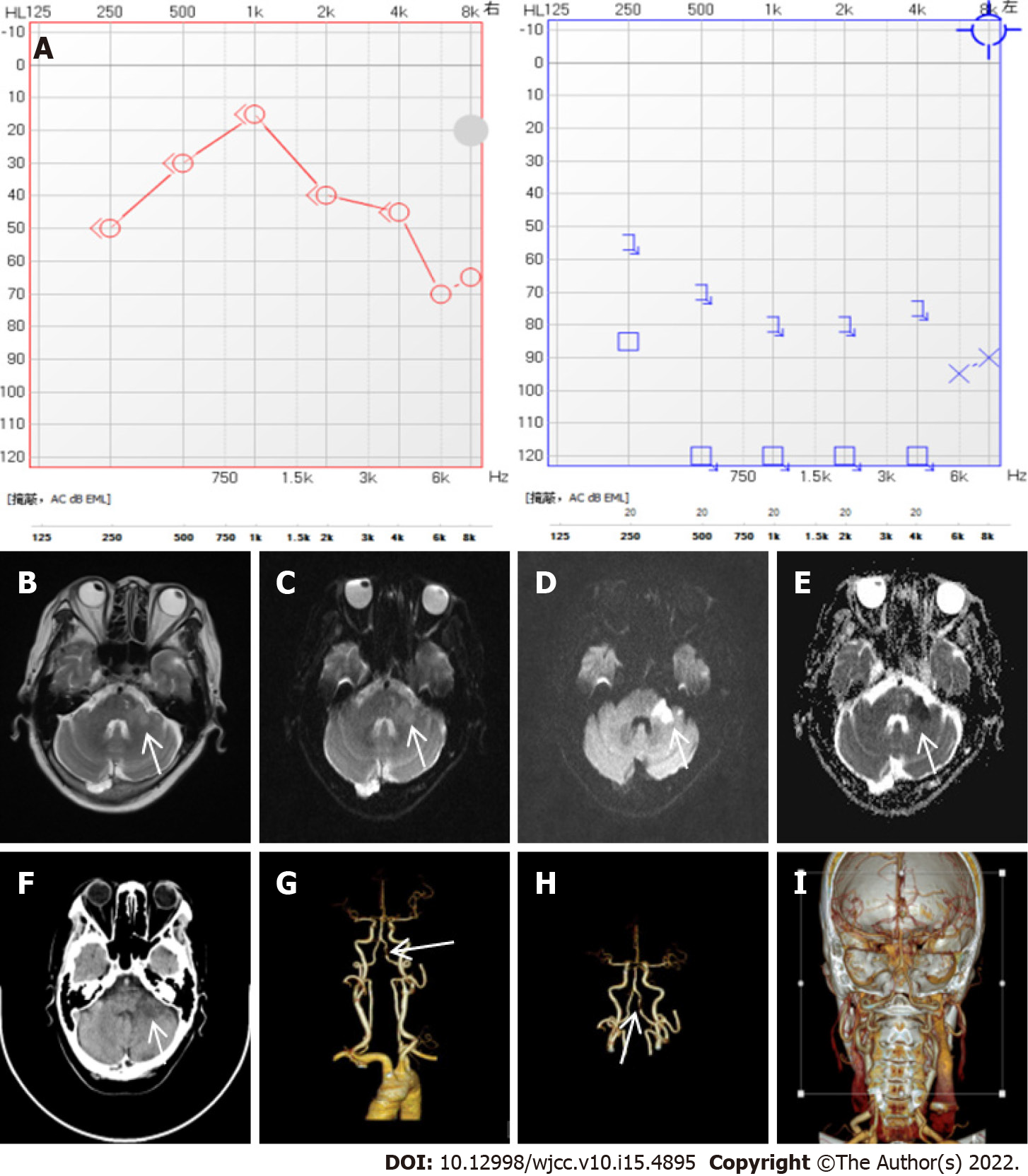
Figure 3 Related auxiliary examination of case three.
A: Pure tone auditory threshold, extremely severe sensorineural deafness in the left ear; B–E: Brain magnetic resonance imaging, patchy and spotted long T1 and long T2 signal shadows were seen in the left midcerebellar foot, and the diffuse image of lesions in the left midcerebellar foot was all high signal; F–I: Brain and neck computed tomography angiography, patchy low-density shadow was seen in the left cerebellum, with slightly blurred edges and a range of about 2.2 cm × 1.6 cm. The initial segment of the left vertebral artery was tortuous and the left vertebral artery V4 segment was moderately and severely narrowed.
- Citation: Li BL, Xu JY, Lin S. Sudden deafness as a prodrome of cerebellar artery infarction: Three case reports. World J Clin Cases 2022; 10(15): 4895-4903
- URL: https://www.wjgnet.com/2307-8960/full/v10/i15/4895.htm
- DOI: https://dx.doi.org/10.12998/wjcc.v10.i15.4895


