Copyright
©The Author(s) 2022.
World J Clin Cases. May 26, 2022; 10(15): 4895-4903
Published online May 26, 2022. doi: 10.12998/wjcc.v10.i15.4895
Published online May 26, 2022. doi: 10.12998/wjcc.v10.i15.4895
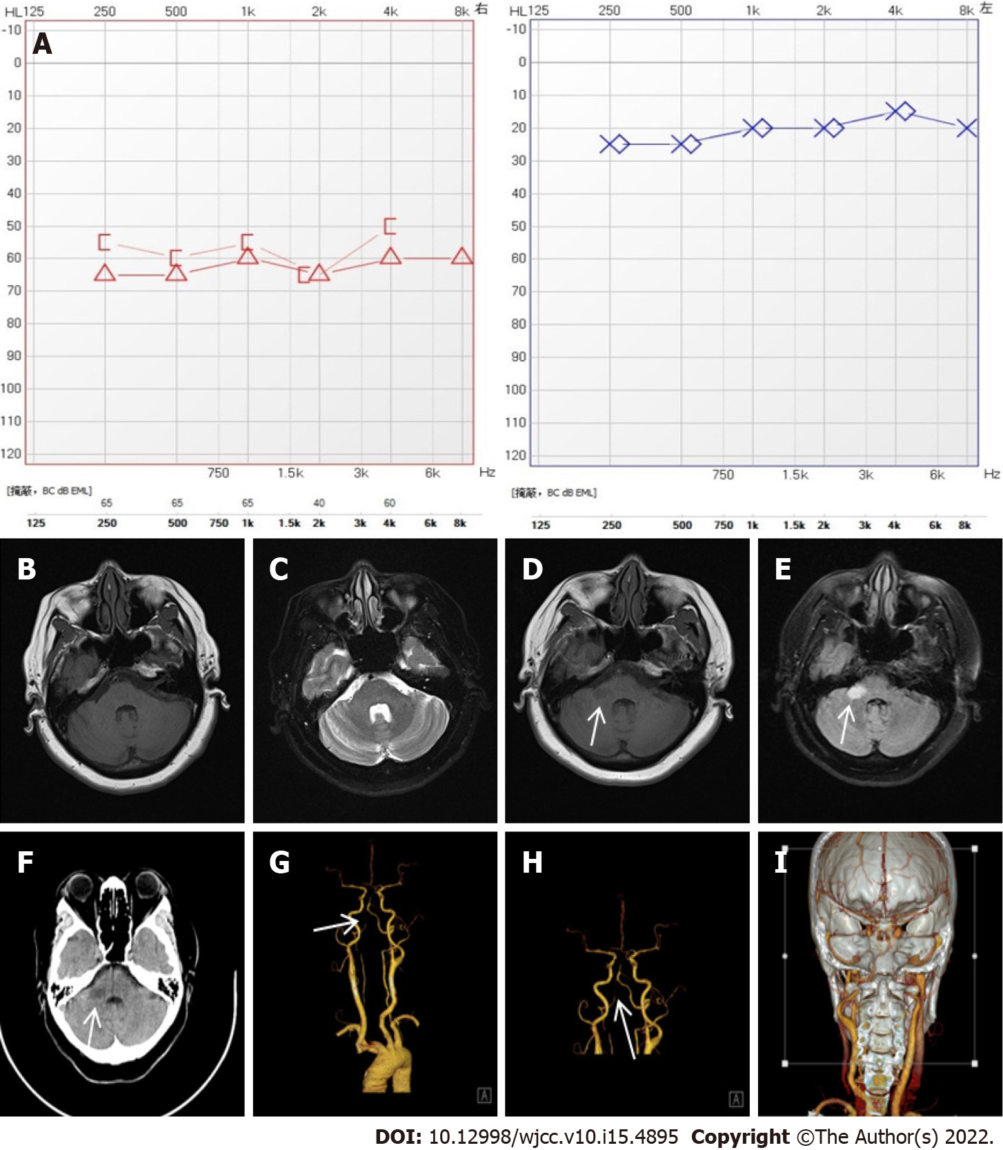
Figure 1 Related auxiliary examination of case one.
A: Pure tone auditory threshold, moderate to severe sensorineural hearing loss in the right ear; B, C: Mastoid magnetic resonance imaging (MRI)(2019-11-12), no obvious abnormalities were found in bilateral internal auditory canal and mastoid process, see pontine lacunar infarction; D, E: Brain MRI (2019-11-19), patchy long T1 and long T2 signal foci were seen in the right cerebellar foot, and diffusion-weighted imaging showed high signal intensity; F–I: Brain and neck computed tomography angiography, patchy low-density shadow was seen in the right cerebellar foot, local lumen stenosis of the right posterior cerebral artery and basilar artery was not clear.
- Citation: Li BL, Xu JY, Lin S. Sudden deafness as a prodrome of cerebellar artery infarction: Three case reports. World J Clin Cases 2022; 10(15): 4895-4903
- URL: https://www.wjgnet.com/2307-8960/full/v10/i15/4895.htm
- DOI: https://dx.doi.org/10.12998/wjcc.v10.i15.4895


