Copyright
©The Author(s) 2022.
World J Clin Cases. May 16, 2022; 10(14): 4704-4708
Published online May 16, 2022. doi: 10.12998/wjcc.v10.i14.4704
Published online May 16, 2022. doi: 10.12998/wjcc.v10.i14.4704
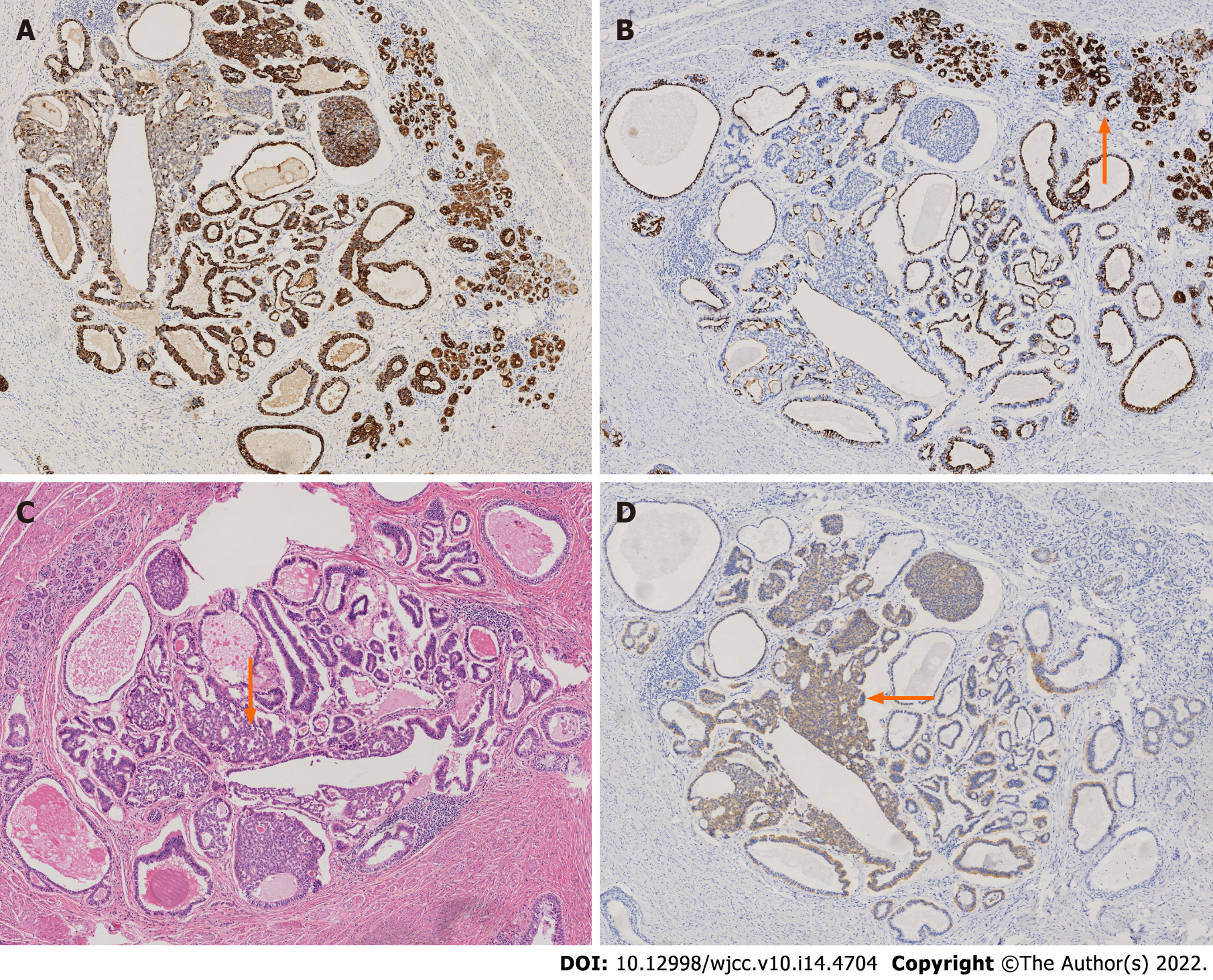
Figure 2 Postoperative histopathology combined with immunohistochemistry analysis demonstrated purely mature teratomatous tissues.
A: Shows that the pathological immunohistochemical results are positive for cytokeratin; B: Shows that the pathological immunohistochemical results are negative for cytokeratin 7; C: Shows that the pathological immunohistochemical results are is HE staining; D: Shows that the pathological immunohistochemical results are positive for cluster of differentiation 56. The pathological results were obtained under a microscope with a magnification of 40 times, in which figure A is cytokeratin (+), figure B is cytokeratin 7 (-), figure C is HE staining, figure D is cluster of differentiation 56 (+), and orange arrows mark a large number of nerves endocrine cell aggregation, and all pathological results showed no immature neural tube, suggesting a mature teratoma.
- Citation: Hu X, Jia Z, Zhou LX, Kakongoma N. Ovarian growing teratoma syndrome with multiple metastases in the abdominal cavity and liver: A case report. World J Clin Cases 2022; 10(14): 4704-4708
- URL: https://www.wjgnet.com/2307-8960/full/v10/i14/4704.htm
- DOI: https://dx.doi.org/10.12998/wjcc.v10.i14.4704


