Copyright
©The Author(s) 2022.
World J Clin Cases. May 16, 2022; 10(14): 4676-4683
Published online May 16, 2022. doi: 10.12998/wjcc.v10.i14.4676
Published online May 16, 2022. doi: 10.12998/wjcc.v10.i14.4676
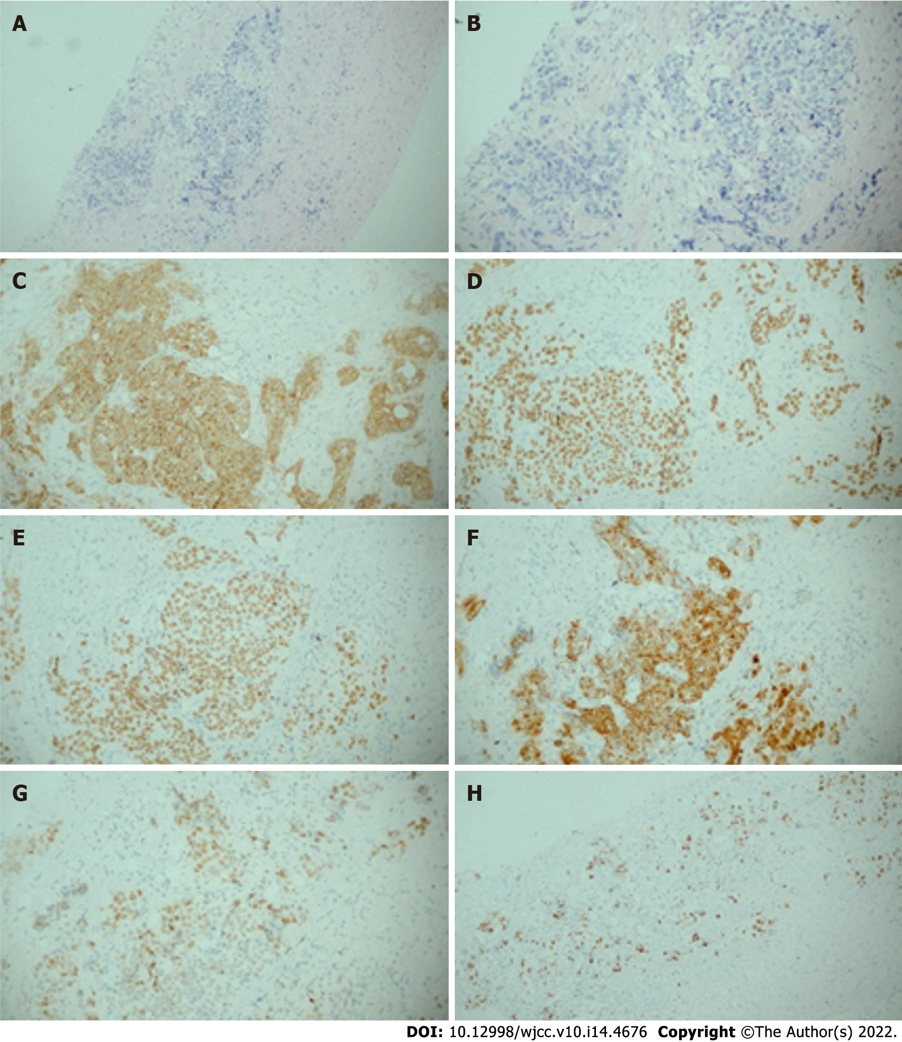
Figure 4 Histological patterns of skin lesions.
A, B: The hematoxylin and eosin staining of sites for the fine needle biopsy specimen revealed esophageal squamous cell carcinoma in A (magnification × 10), B (magnification × 20); C: Representative immunohistochemical staining for CK in the skin (magnification × 20); D:: Representative immunohistochemical staining for P40 in the skin (magnification × 20); E: Representative immunohistochemical staining for P63 in the skin (magnification × 20); F: Representative immunohistochemical staining for CK5/6 in the skin (magnification × 20); G: Representative immunohistochemical staining for P53 in the skin (magnification × 20); H: Representative immunohistochemical staining for Ki67 in the skin (magnification × 10).
- Citation: Zhang RY, Zhu SJ, Xue P, He SQ. Cutaneous metastasis from esophageal squamous cell carcinoma: A case report. World J Clin Cases 2022; 10(14): 4676-4683
- URL: https://www.wjgnet.com/2307-8960/full/v10/i14/4676.htm
- DOI: https://dx.doi.org/10.12998/wjcc.v10.i14.4676


