Copyright
©The Author(s) 2022.
World J Clin Cases. May 16, 2022; 10(14): 4676-4683
Published online May 16, 2022. doi: 10.12998/wjcc.v10.i14.4676
Published online May 16, 2022. doi: 10.12998/wjcc.v10.i14.4676
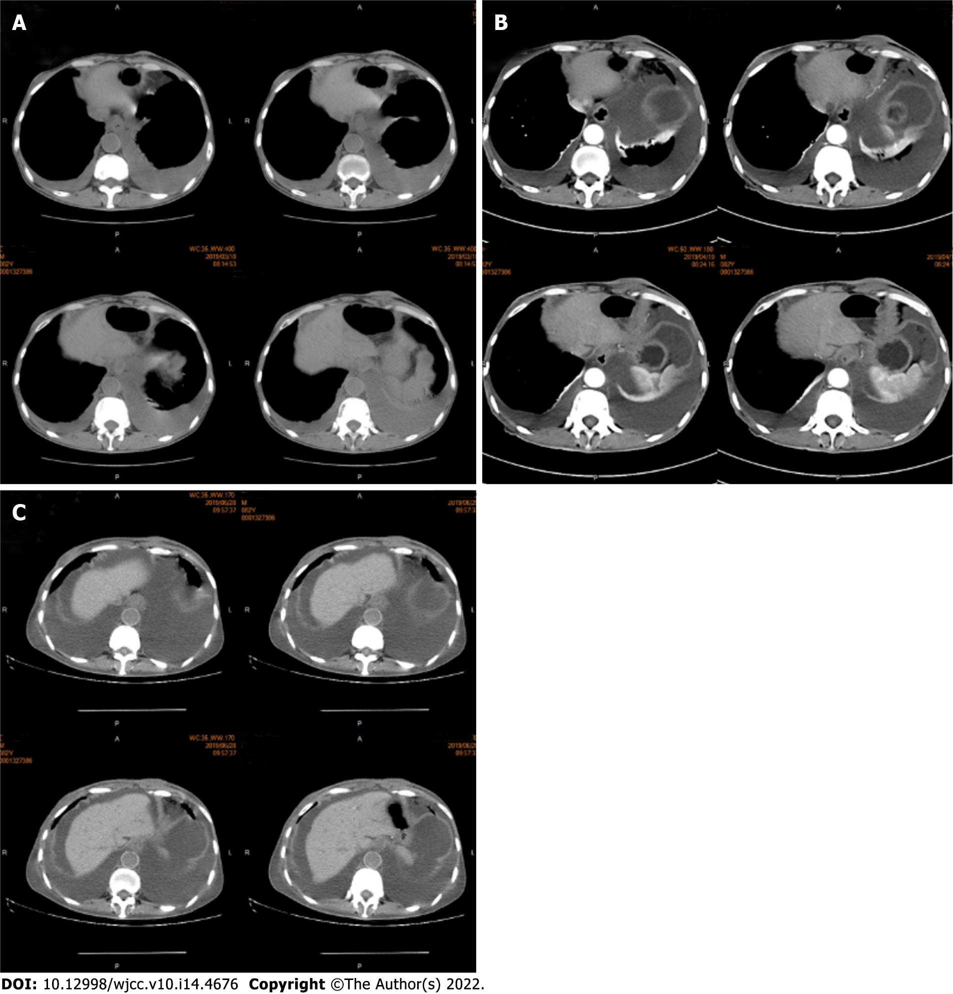
Figure 3 Abdominal computed tomography of the skin and subcutaneous soft tissue.
A: The chest computed tomography presented pleural effusion, suggesting that the disease recurred; B: This suggested ascites effusion as well as lower esophagus dilation and esophageal wall thickening; C: After two cycles of nivolumab treatment, the effusion increased, and the lower esophagus thickening became obvious. The abdominal computed tomography showed that the abdominal wall skin was thicker than the other parts, but no subcutaneous nodules could be observed. Imaging diagnosis can easily overlook skin thickening.
- Citation: Zhang RY, Zhu SJ, Xue P, He SQ. Cutaneous metastasis from esophageal squamous cell carcinoma: A case report. World J Clin Cases 2022; 10(14): 4676-4683
- URL: https://www.wjgnet.com/2307-8960/full/v10/i14/4676.htm
- DOI: https://dx.doi.org/10.12998/wjcc.v10.i14.4676


