Copyright
©The Author(s) 2022.
World J Clin Cases. May 16, 2022; 10(14): 4625-4631
Published online May 16, 2022. doi: 10.12998/wjcc.v10.i14.4625
Published online May 16, 2022. doi: 10.12998/wjcc.v10.i14.4625
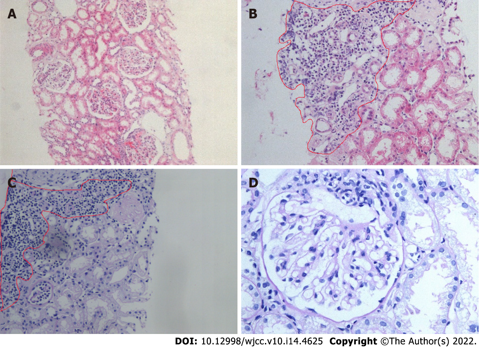
Figure 1 Kidney histological examination.
A: Hematoxylin-eosin staining, ×20; B: Hematoxylin-eosin staining, ×20; C: Periodic acid-Schiff stain, ×20; D: Periodic acid-Schiff stain, ×40; B and C: Light microscopic analysis of a kidney biopsy sample showed no abnormalities in the glomeruli and occasional focal aggregation of interstitial inflammatory cells.
- Citation: Li CY, Li YM, Tian M. Serum-negative Sjogren's syndrome with minimal lesion nephropathy as the initial presentation: A case report. World J Clin Cases 2022; 10(14): 4625-4631
- URL: https://www.wjgnet.com/2307-8960/full/v10/i14/4625.htm
- DOI: https://dx.doi.org/10.12998/wjcc.v10.i14.4625


