Copyright
©The Author(s) 2022.
World J Clin Cases. May 16, 2022; 10(14): 4617-4624
Published online May 16, 2022. doi: 10.12998/wjcc.v10.i14.4617
Published online May 16, 2022. doi: 10.12998/wjcc.v10.i14.4617
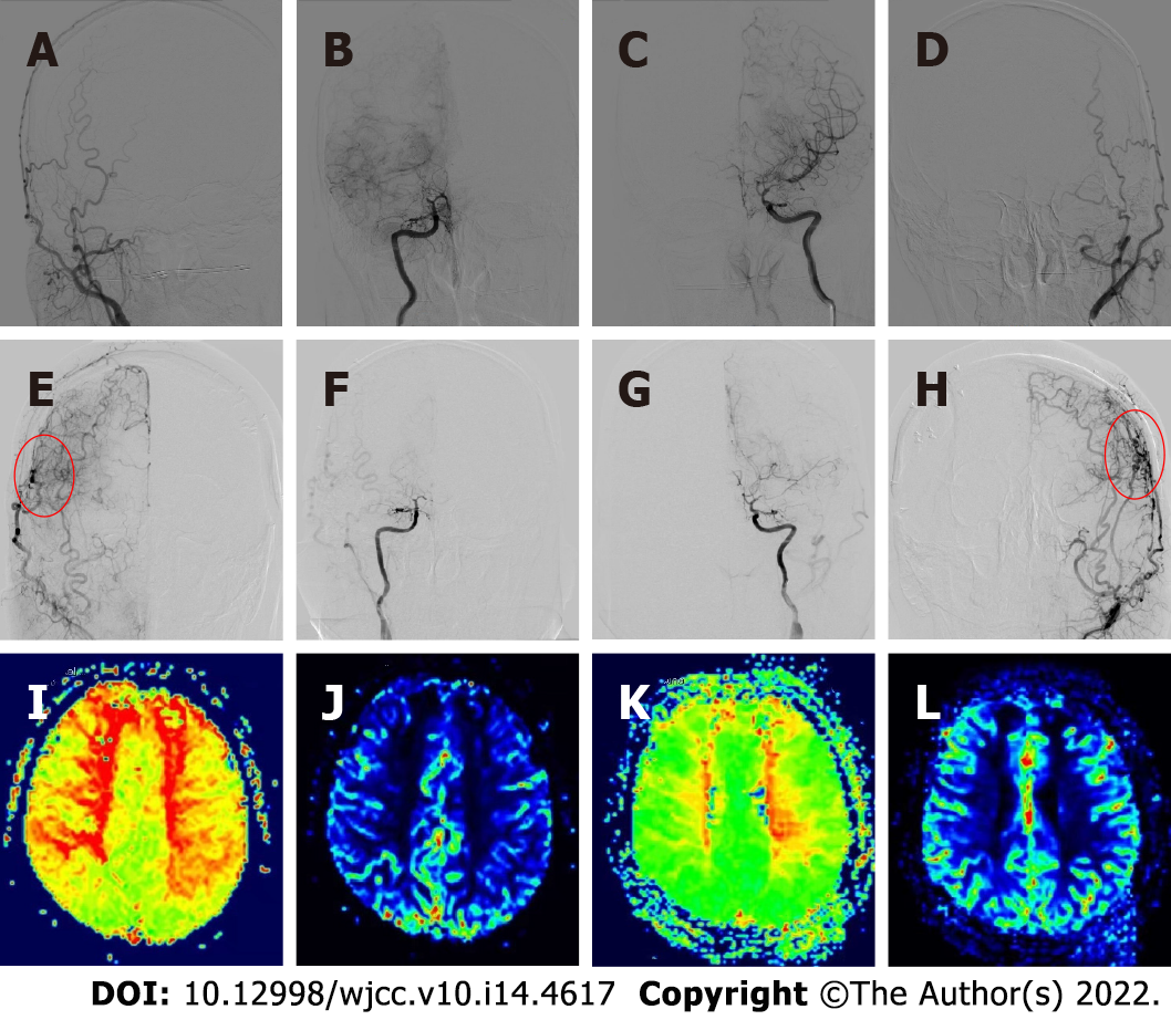
Figure 2 Preoperative and postoperative angiography and cerebral perfusion contrast.
A-D: Preoperative digital subtraction angiography showed moyamoya disease; E-H: The neovascularization was good after bilateral combined cerebral revascularization; I and J: Preoperative magnetic resonance-perfusion-weighted imaging (MR-PWI) showed prolonged time to peak and decreased regional cerebral blood flow in the bilateral frontal parietal lobe, bilateral paraventricular region, and center of semicovale; K and L: Six months after bilateral cerebral revascularization, MR-PWI showed significant improvement in bilateral cerebral perfusion.
- Citation: Zhang S, Zhao LM, Xue BQ, Liang H, Guo GC, Liu Y, Wu RY, Li CY. Acute recurrent cerebral infarction caused by moyamoya disease complicated with adenomyosis: A case report . World J Clin Cases 2022; 10(14): 4617-4624
- URL: https://www.wjgnet.com/2307-8960/full/v10/i14/4617.htm
- DOI: https://dx.doi.org/10.12998/wjcc.v10.i14.4617


