Copyright
©The Author(s) 2022.
World J Clin Cases. May 16, 2022; 10(14): 4541-4549
Published online May 16, 2022. doi: 10.12998/wjcc.v10.i14.4541
Published online May 16, 2022. doi: 10.12998/wjcc.v10.i14.4541
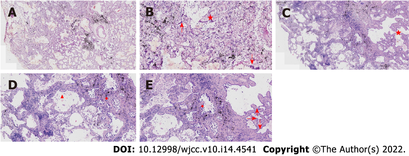
Figure 4 Pathological features of case 2.
A: At low magnification (100 ×, frozen section), the boundary of the tumor was relatively clear; there were air cavities and arterioles were visible; B: At high magnification (200 ×, frozen section), the tumor cells were found to be mainly arranged in a monolayer with locally visible cilia (arrows); some nuclei appeared enlarged and atypical (star); C: At low magnification (100 ×), most cells appeared with moderate density (star), focal hyperplasia, and stroma within the focal lymphocytic infiltration; D and E: Observations at medium to high magnification (200 × and 400 ×, respectively) revealed that the tumor cells were arranged as an acinar structure and accessory wall structure; most cells were not atypia in shape, and cilia (arrow) were seen. Some nuclei were enlarged and atypical (circle).
- Citation: Du Y, Wang ZY, Zheng Z, Li YX, Wang XY, Du R. Bronchiolar adenoma with unusual presentation: Two case reports. World J Clin Cases 2022; 10(14): 4541-4549
- URL: https://www.wjgnet.com/2307-8960/full/v10/i14/4541.htm
- DOI: https://dx.doi.org/10.12998/wjcc.v10.i14.4541


