Copyright
©The Author(s) 2022.
World J Clin Cases. May 16, 2022; 10(14): 4494-4501
Published online May 16, 2022. doi: 10.12998/wjcc.v10.i14.4494
Published online May 16, 2022. doi: 10.12998/wjcc.v10.i14.4494
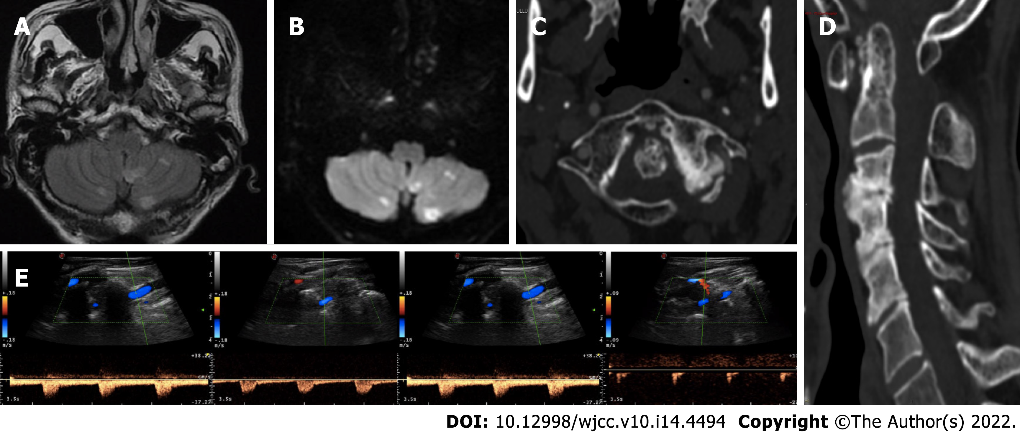
Figure 1 Neuroradiological and neurosonological evaluation.
A and B: Axial fluid-attenuated inversion-recovery and diffusion-weighted imaging (DWI) brain magnetic resonance imaging sequences with subacute bilateral cerebellar ischaemic lesions involving the area supplied by the posterior inferior cerebellar artery; C and D: Axial and sagittal cervical computed tomography scan showing marked degenerative joint alterations with atlo-axial instability and retroposition of dens, spinal canal stenosis and ankilosis of lateral left zygo-apophisal joints with underlying congenital partial atlo-occipital fusion; E: Vertebral ultrasound examination documenting regular blood flow in the left VA in the neutral position (left side) and “stump flow” demodulation in both the V1-V2 and V3-V4 segments (right side) in cases of slight contralateral head rotation (20°).
- Citation: Orlandi N, Cavallieri F, Grisendi I, Romano A, Ghadirpour R, Napoli M, Moratti C, Zanichelli M, Pascarella R, Valzania F, Zedde M. Bow hunter’s syndrome successfully treated with a posterior surgical decompression approach: A case report and review of literature. World J Clin Cases 2022; 10(14): 4494-4501
- URL: https://www.wjgnet.com/2307-8960/full/v10/i14/4494.htm
- DOI: https://dx.doi.org/10.12998/wjcc.v10.i14.4494


