Copyright
©The Author(s) 2022.
World J Clin Cases. May 16, 2022; 10(14): 4436-4445
Published online May 16, 2022. doi: 10.12998/wjcc.v10.i14.4436
Published online May 16, 2022. doi: 10.12998/wjcc.v10.i14.4436
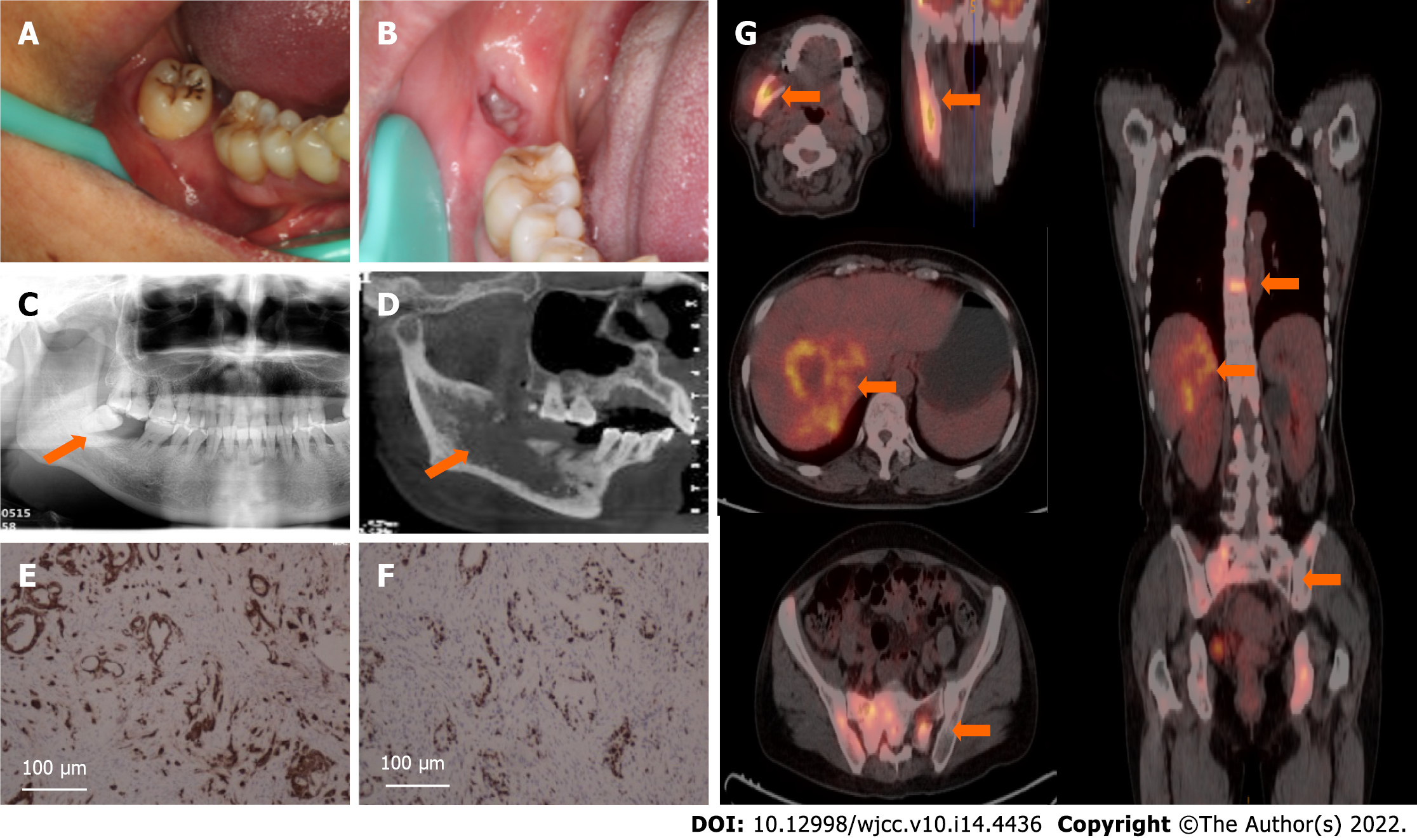
Figure 4 A patient with metastatic adenocarcinoma of the jaw of primary liver cancer.
A: Intraoral image before tooth extraction showed swelling of the gingival surrounding the wisdom tooth (arrow); B: Panorama radiograph before tooth extraction showed periodontal bone loss around the involved tooth (arrow); C: At a two-month follow-up visit, the extraction wound did not heal completely; D: Four months after tooth extraction, oblique sagittal cone beam computed tomography (CT) showed lesion (arrow) turned bulky with a permeative margin. The wall of the inferior alveolar nerve was invisible; E: Immunohistochemistry for the expression of CK8/18 revealed uniform positivity in the cytoplasm of tumour cells (Magnification: 100 ×); F: Immunohistochemistry for the expression of Ki-67 revealed scattered positivity in more than 65% of the tumour cells (Magnification: 100 ×); G: Positron emission tomography-CT scans showed asymptomatic hepatocellular carcinoma as the primary site and multiple metastases mainly involving the right mandible, spine, and bilateral pelvic bone (arrows).
- Citation: Shan S, Liu S, Yang ZY, Wang TM, Lin ZT, Feng YL, Pakezhati S, Huang XF, Zhang L, Sun GW. Oral and maxillofacial pain as the first sign of metastasis of an occult primary tumour: A fifteen-year retrospective study. World J Clin Cases 2022; 10(14): 4436-4445
- URL: https://www.wjgnet.com/2307-8960/full/v10/i14/4436.htm
- DOI: https://dx.doi.org/10.12998/wjcc.v10.i14.4436


