Copyright
©The Author(s) 2022.
World J Clin Cases. May 6, 2022; 10(13): 4314-4320
Published online May 6, 2022. doi: 10.12998/wjcc.v10.i13.4314
Published online May 6, 2022. doi: 10.12998/wjcc.v10.i13.4314
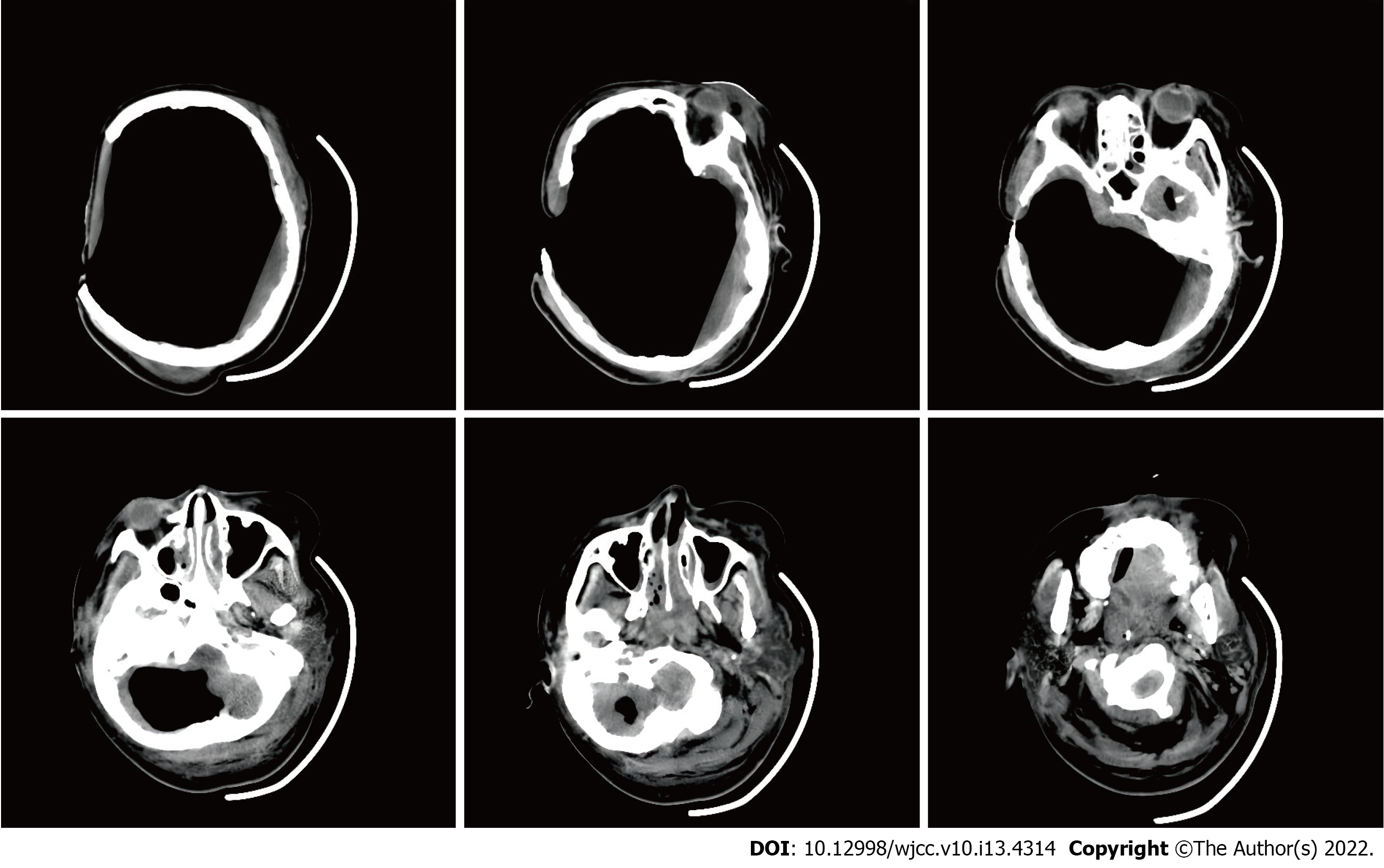
Figure 3 Cranial computed tomography.
A large amount of gas was visualized in the intracranial and paranasal sinuses, and only a small amount of brain tissue density shadow was compressed in the cerebellum and brainstem. Moreover, the sulcus, fissure, cistern, ventricle, inferior mourning, and midline structures were not shown.
- Citation: Li GG, Zhang ZQ, Mi YH. Mass brain tissue lost after decompressive craniectomy: A case report. World J Clin Cases 2022; 10(13): 4314-4320
- URL: https://www.wjgnet.com/2307-8960/full/v10/i13/4314.htm
- DOI: https://dx.doi.org/10.12998/wjcc.v10.i13.4314


