Copyright
©The Author(s) 2022.
World J Clin Cases. May 6, 2022; 10(13): 4294-4300
Published online May 6, 2022. doi: 10.12998/wjcc.v10.i13.4294
Published online May 6, 2022. doi: 10.12998/wjcc.v10.i13.4294
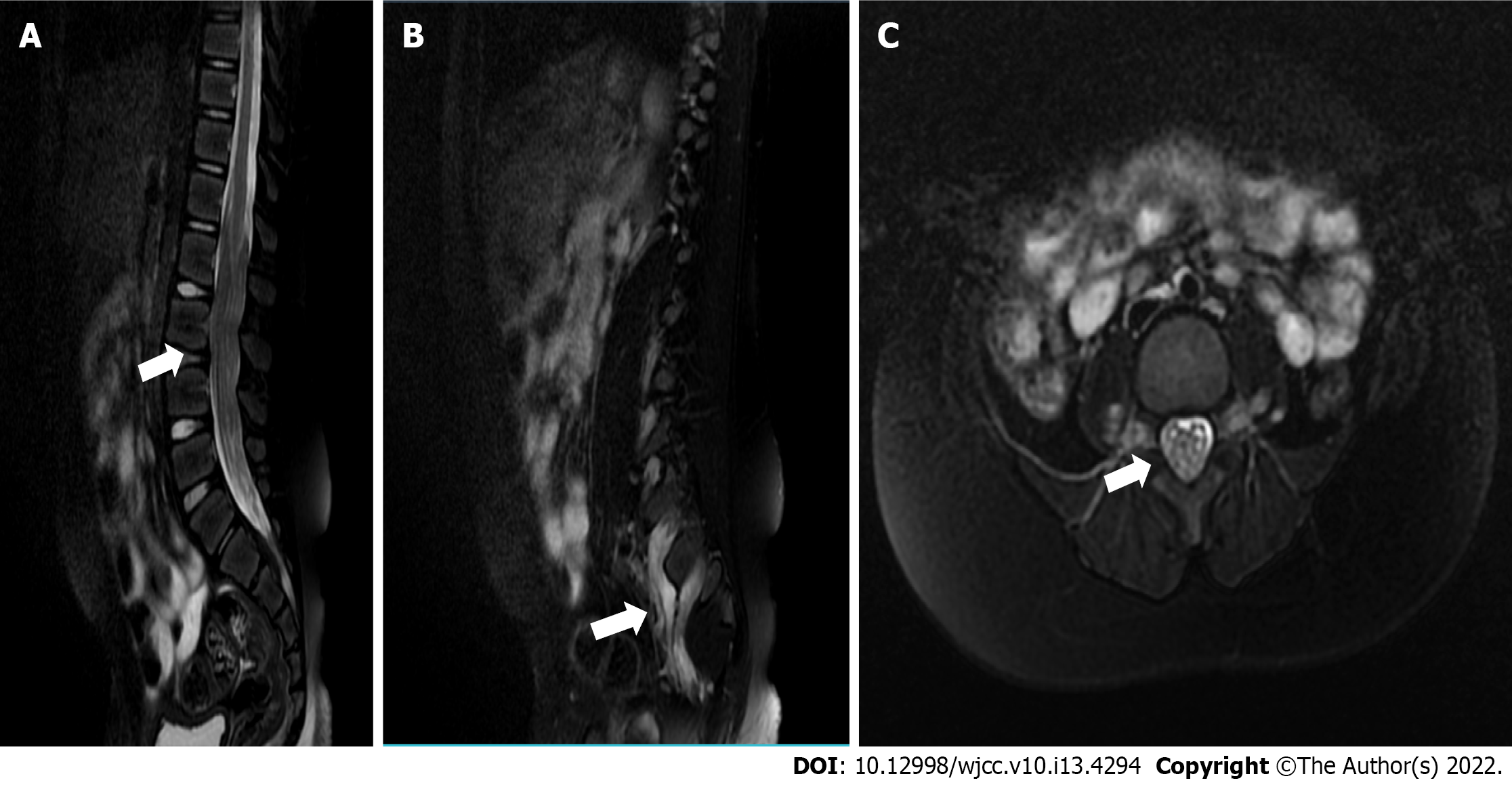
Figure 1 Image of T2-weighted magnetic resonance imaging.
A: Diffuse thickening of nerve roots in the spinal canal is seen on magnetic resonance imaging (MRI) sagittal T2 slices (white arrow); B: Extraforaminal nerve root also showed signs of thickening on MRI sagittal T2 slices (white arrow); C: The spinal canal was almost filled with hypertrophic cauda equina on MRI axial T2 slices (white arrow).
- Citation: Ye L, Yu W, Liang NZ, Sun Y, Duan LF. Spinal canal decompression for hypertrophic neuropathy of the cauda equina with chronic inflammatory demyelinating polyradiculoneuropathy: A case report. World J Clin Cases 2022; 10(13): 4294-4300
- URL: https://www.wjgnet.com/2307-8960/full/v10/i13/4294.htm
- DOI: https://dx.doi.org/10.12998/wjcc.v10.i13.4294


