Copyright
©The Author(s) 2022.
World J Clin Cases. May 6, 2022; 10(13): 4280-4287
Published online May 6, 2022. doi: 10.12998/wjcc.v10.i13.4280
Published online May 6, 2022. doi: 10.12998/wjcc.v10.i13.4280
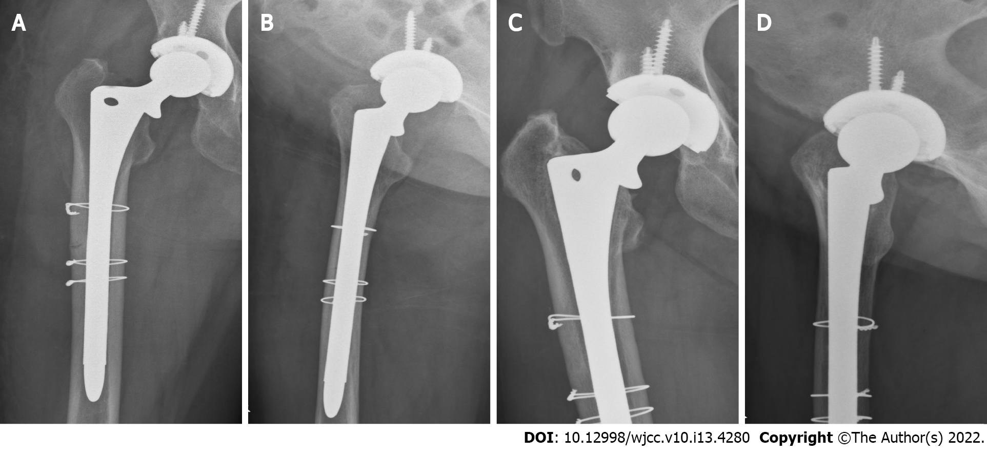
Figure 4 X-rays of hip joint after the operation and 3 mo after the operation.
A: Anterior-posterior view; B: Lateral view. Both views show that, after total hip arthroplasty, the prosthesis was in a good position and the proximal femur was fixed with steel wires to prevent intraoperative fracture; C: Anterior-posterior view; D: Lateral view. Both views show that the right artificial hip prosthesis was in a good position, the fracture line of the right proximal femur disappeared, and the bone fracture healed well.
- Citation: Tang MT, Liu CF, Liu JL, Saijilafu, Wang Z. Multiple stress fractures of unilateral femur: A case report . World J Clin Cases 2022; 10(13): 4280-4287
- URL: https://www.wjgnet.com/2307-8960/full/v10/i13/4280.htm
- DOI: https://dx.doi.org/10.12998/wjcc.v10.i13.4280


