Copyright
©The Author(s) 2022.
World J Clin Cases. May 6, 2022; 10(13): 4280-4287
Published online May 6, 2022. doi: 10.12998/wjcc.v10.i13.4280
Published online May 6, 2022. doi: 10.12998/wjcc.v10.i13.4280
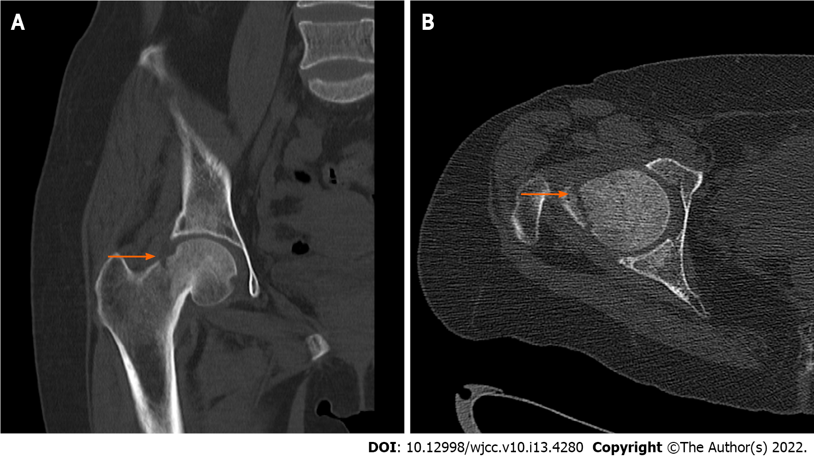
Figure 3 Preoperative computed tomography of the hip joint.
A: Coronal scan; B: Axial scan. Both scans show the fracture line above the femoral neck (arrow), obvious displacement of the fracture end, and varus deformity of the hip.
- Citation: Tang MT, Liu CF, Liu JL, Saijilafu, Wang Z. Multiple stress fractures of unilateral femur: A case report . World J Clin Cases 2022; 10(13): 4280-4287
- URL: https://www.wjgnet.com/2307-8960/full/v10/i13/4280.htm
- DOI: https://dx.doi.org/10.12998/wjcc.v10.i13.4280


