Copyright
©The Author(s) 2022.
World J Clin Cases. May 6, 2022; 10(13): 4280-4287
Published online May 6, 2022. doi: 10.12998/wjcc.v10.i13.4280
Published online May 6, 2022. doi: 10.12998/wjcc.v10.i13.4280
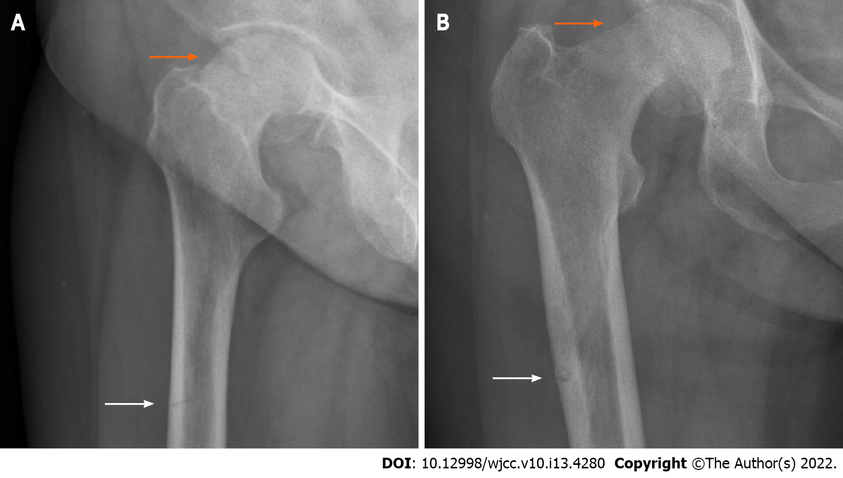
Figure 2 Plain radiograph of the hip joint before an operation.
A: Lateral view; B: Anterior-posterior view. Both show the fracture line at the upper end of the right femoral neck (orange arrow), displacement of the fracture end, varus deformity of the hip, and proximal femur cortical discontinuity (white arrow).
- Citation: Tang MT, Liu CF, Liu JL, Saijilafu, Wang Z. Multiple stress fractures of unilateral femur: A case report . World J Clin Cases 2022; 10(13): 4280-4287
- URL: https://www.wjgnet.com/2307-8960/full/v10/i13/4280.htm
- DOI: https://dx.doi.org/10.12998/wjcc.v10.i13.4280


