Copyright
©The Author(s) 2022.
World J Clin Cases. May 6, 2022; 10(13): 4273-4279
Published online May 6, 2022. doi: 10.12998/wjcc.v10.i13.4273
Published online May 6, 2022. doi: 10.12998/wjcc.v10.i13.4273
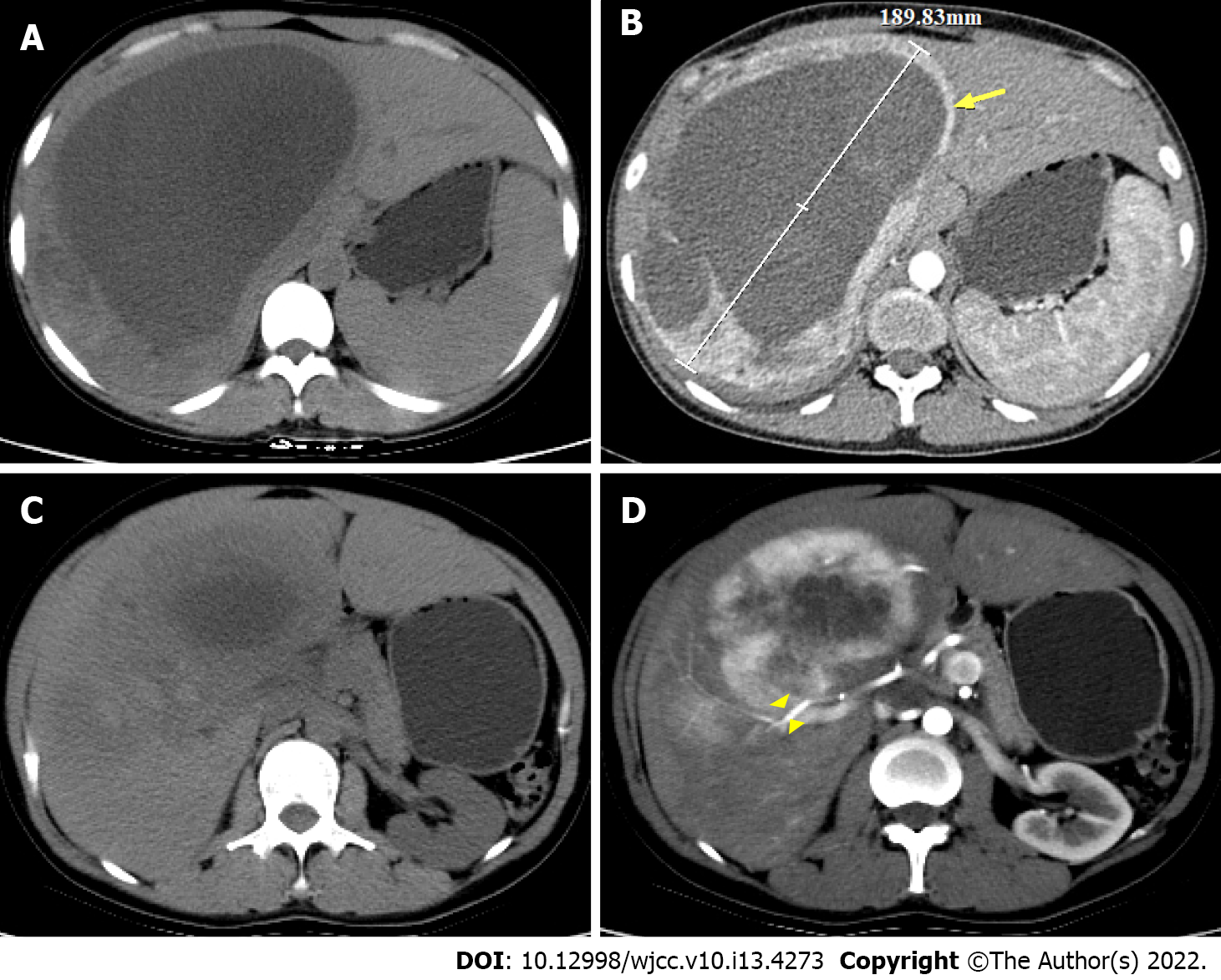
Figure 1 Abdominal computed tomography scan findings (axial).
A-C: Large oval cystic solid space-occupying lesion, maximum diameter was approximately 18.9 cm in S7 and S8 segments of the liver; B and D: Solid component of the tumor (arrow) in arterial phase showing heterogeneous and marked enhancement, the cystic components had no obvious enhancement effect, with penetration by hepatic artery branches figure (arrowhead).
- Citation: Li YF, Wang L, Xie YJ. Hepatic perivascular epithelioid cell tumor: A case report. World J Clin Cases 2022; 10(13): 4273-4279
- URL: https://www.wjgnet.com/2307-8960/full/v10/i13/4273.htm
- DOI: https://dx.doi.org/10.12998/wjcc.v10.i13.4273


