Copyright
©The Author(s) 2022.
World J Clin Cases. May 6, 2022; 10(13): 4264-4272
Published online May 6, 2022. doi: 10.12998/wjcc.v10.i13.4264
Published online May 6, 2022. doi: 10.12998/wjcc.v10.i13.4264
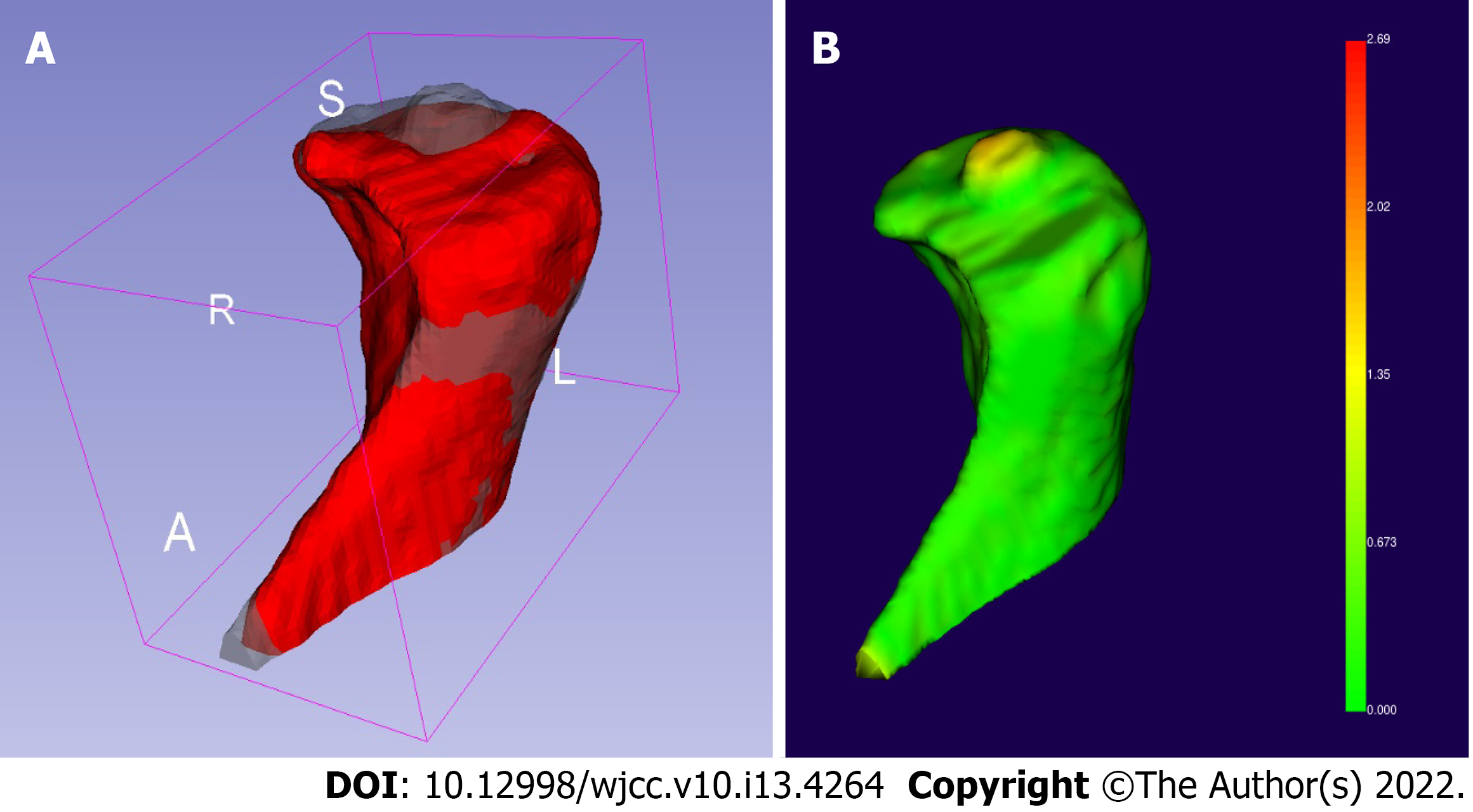
Figure 5 Three-dimensional reconstruction of cone beam computed tomography radiographs.
A: The reconstruction models of the left condyle before treatment (gray model) and 9 mo after treatment (red model) were compared using 3D Slicer version 4.10.2 (https://download.slicer.org); B: We calculated the facial distance of the registration model and found that the 2-mm-high cylindrical osteophyte on top of the condyle had dissolved.
- Citation: Lan KW, Chen JM, Jiang LL, Feng YF, Yan Y. Treatment of condylar osteophyte in temporomandibular joint osteoarthritis with muscle balance occlusal splint and long-term follow-up: A case report. World J Clin Cases 2022; 10(13): 4264-4272
- URL: https://www.wjgnet.com/2307-8960/full/v10/i13/4264.htm
- DOI: https://dx.doi.org/10.12998/wjcc.v10.i13.4264


