Copyright
©The Author(s) 2022.
World J Clin Cases. May 6, 2022; 10(13): 4226-4235
Published online May 6, 2022. doi: 10.12998/wjcc.v10.i13.4226
Published online May 6, 2022. doi: 10.12998/wjcc.v10.i13.4226
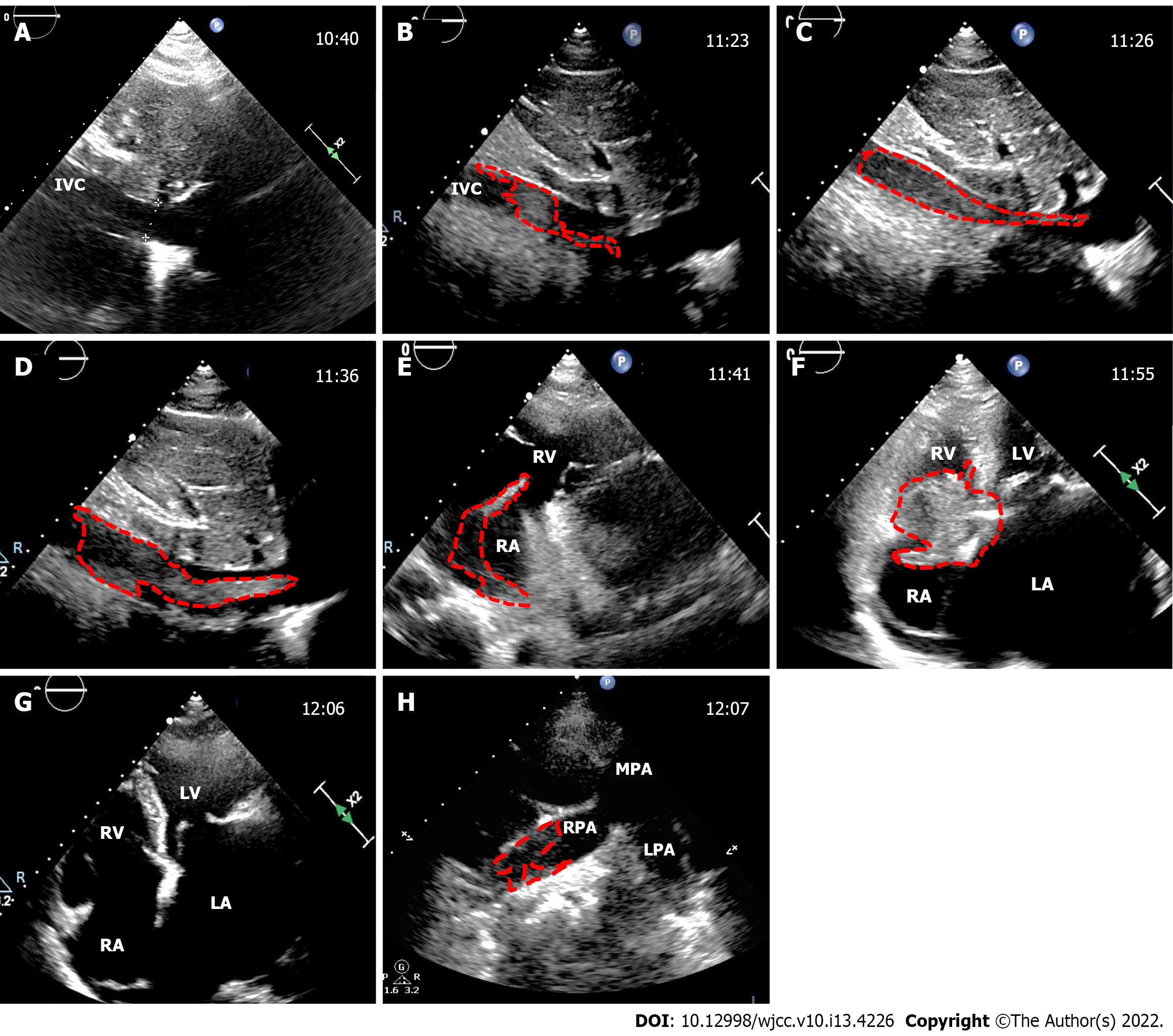
Figure 1 The dynamic process of inferior vena cava thrombus.
The images obtained from transthoracic echocardiography during caesarean section (CS); the time is displayed in the upper left corner; the dotted line outlines the thrombus. A: Inferior vena cava (IVC) was normal before CS; B: A nascent IVC thrombus appeared after placental removal during CS; C-E: The thrombus grew rapidly and extended to the tricuspid valve orifice in a serpentine manner; F: The thrombus moved to the tricuspid valve orifice; G: The thrombus detached from the heart; H: A partial thrombus was seen in the right main pulmonary artery. IVC: Inferior vena cava; LA: Left atrium; RA: Right atrium; LV: Left ventricle; RV: Right ventricle; RPA: Right pulmonary artery; LPA: Left pulmonary artery; MPA: Main pulmonary artery.
- Citation: Jiang L, Liang WX, Yan Y, Wang SP, Dai L, Chen DJ. Thrombotic pulmonary embolism of inferior vena cava during caesarean section: A case report and review of the literature. World J Clin Cases 2022; 10(13): 4226-4235
- URL: https://www.wjgnet.com/2307-8960/full/v10/i13/4226.htm
- DOI: https://dx.doi.org/10.12998/wjcc.v10.i13.4226


