Copyright
©The Author(s) 2022.
World J Clin Cases. May 6, 2022; 10(13): 4020-4032
Published online May 6, 2022. doi: 10.12998/wjcc.v10.i13.4020
Published online May 6, 2022. doi: 10.12998/wjcc.v10.i13.4020
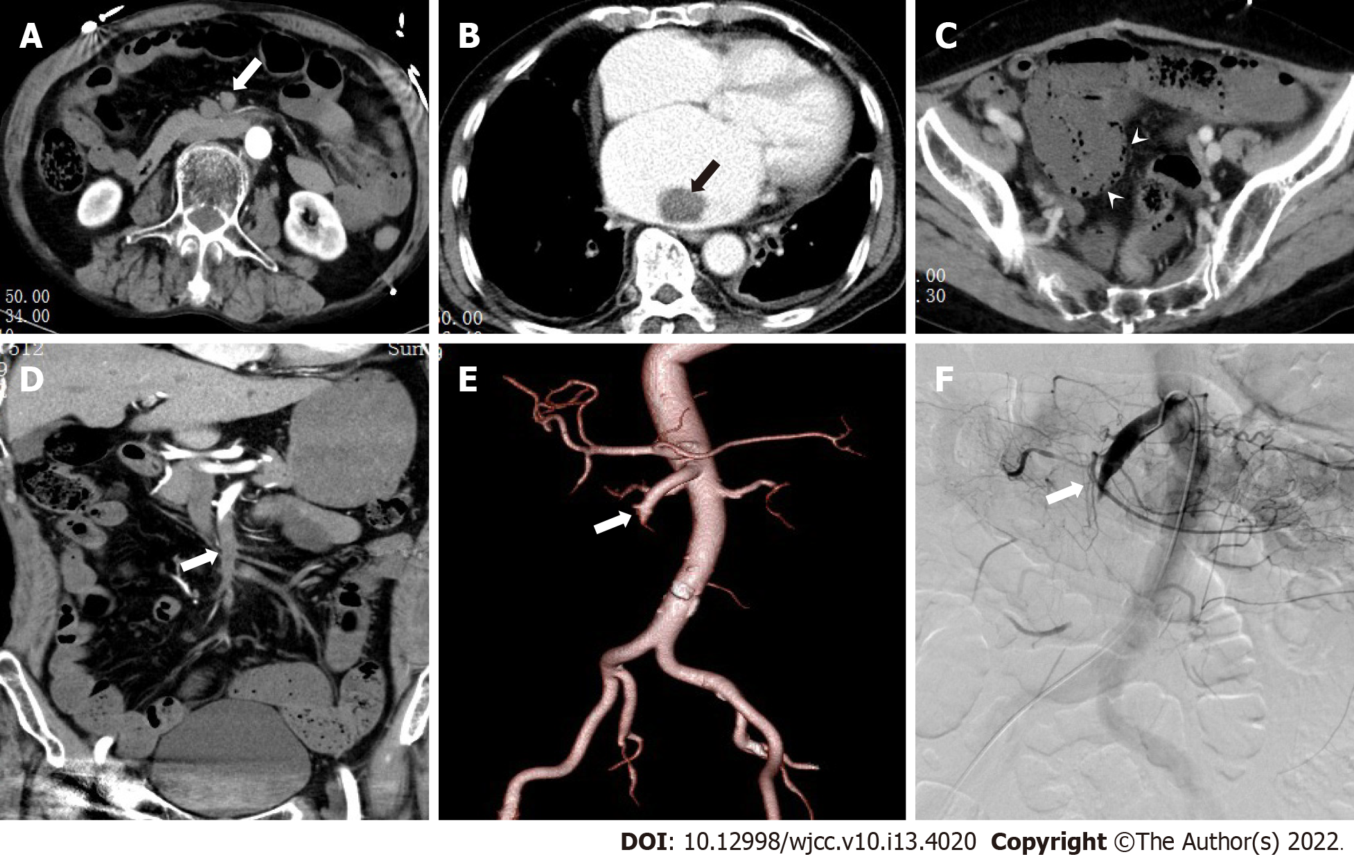
Figure 3 The patient was a 62-year-old male with history of abdominal pain, hematochezia, and atrial fibrillation.
A and B: The axial images in the arterial phase; C: The axial images in the venous phase; D: The coronary images in the arterial phase; E: The volume rendered technique image; and F: The digital subtraction image. A and D show diffuse embolism in II, III, and IV regions of superior mesenteric artery (long arrows); B shows embolus in the left atrium, C shows decreased intestinal wall enhancement, and signs of pneumatosis intestinalis (arrow).
- Citation: Yang JS, Xu ZY, Chen FX, Wang MR, Cong RC, Fan XL, He BS, Xing W. Role of clinical data and multidetector computed tomography findings in acute superior mesenteric artery embolism. World J Clin Cases 2022; 10(13): 4020-4032
- URL: https://www.wjgnet.com/2307-8960/full/v10/i13/4020.htm
- DOI: https://dx.doi.org/10.12998/wjcc.v10.i13.4020


