Copyright
©The Author(s) 2022.
World J Clin Cases. Apr 26, 2022; 10(12): 3936-3943
Published online Apr 26, 2022. doi: 10.12998/wjcc.v10.i12.3936
Published online Apr 26, 2022. doi: 10.12998/wjcc.v10.i12.3936
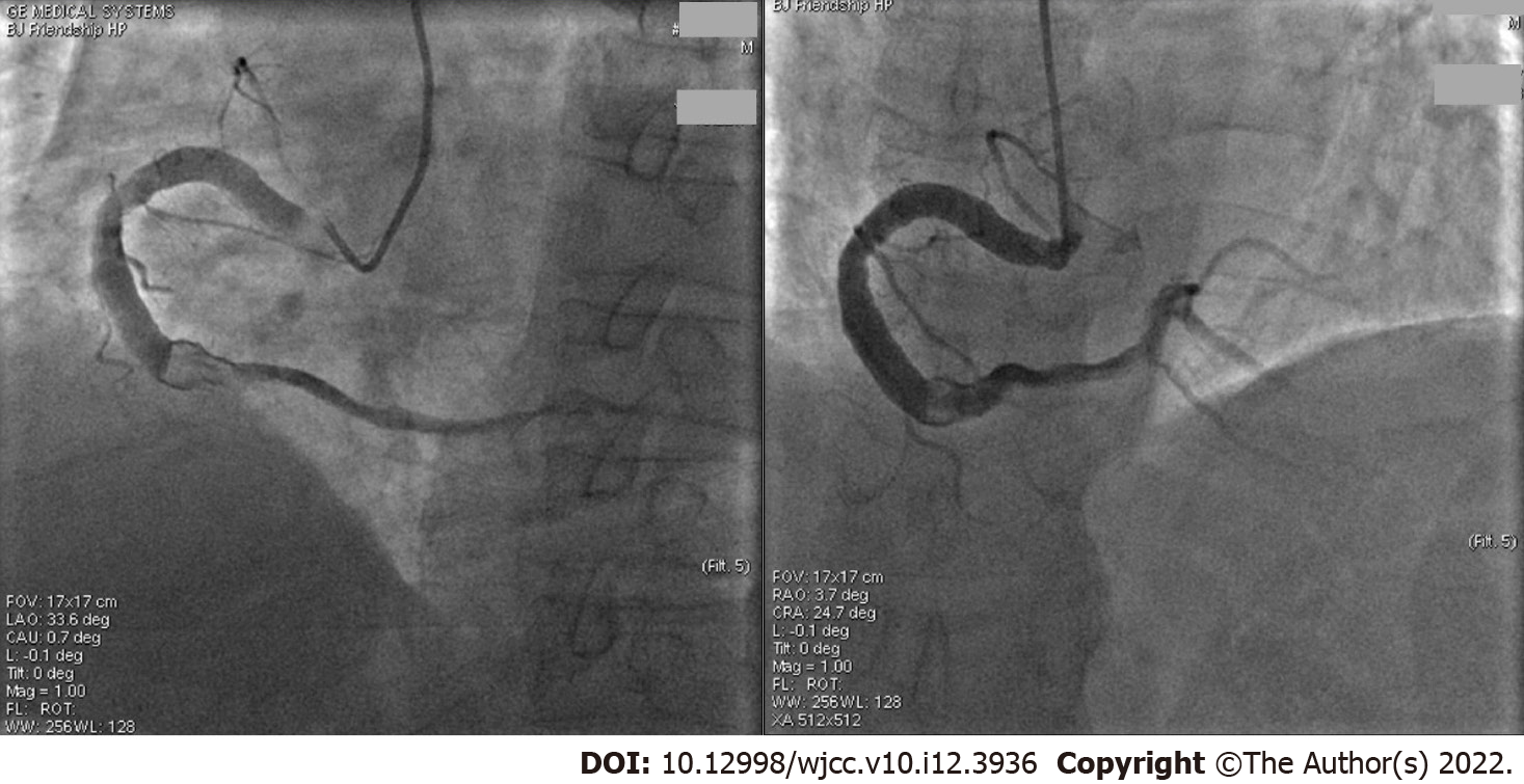
Figure 1 The first coronary angiography.
This angiography was performed 3 d after admission to hospital with chest pain (video), Q wave on electrocardiogram, and increased myocardial injury markers. The two images show aneurysmal dilation in the entire right coronary artery (RCA) with a diameter of 6.60 mm, four fold of a 5 French catheter and more than 1.70 fold of the normal RCA. The middle of the RCA presented with 70%-80% limited stenosis, showed a thrombus shadow after the second turning point, and the forward blood flow was thrombolysis in myocardial infarction grade 3. No additional percutaneous coronary intervention (PCI) was performed after a discussion with the PCI group because the symptoms were relieved and the coronary blood flow was unobstructed.
- Citation: Liu RF, Gao XY, Liang SW, Zhao HQ. Antithrombotic treatment strategy for patients with coronary artery ectasia and acute myocardial infarction: A case report. World J Clin Cases 2022; 10(12): 3936-3943
- URL: https://www.wjgnet.com/2307-8960/full/v10/i12/3936.htm
- DOI: https://dx.doi.org/10.12998/wjcc.v10.i12.3936


