Copyright
©The Author(s) 2022.
World J Clin Cases. Apr 26, 2022; 10(12): 3907-3915
Published online Apr 26, 2022. doi: 10.12998/wjcc.v10.i12.3907
Published online Apr 26, 2022. doi: 10.12998/wjcc.v10.i12.3907
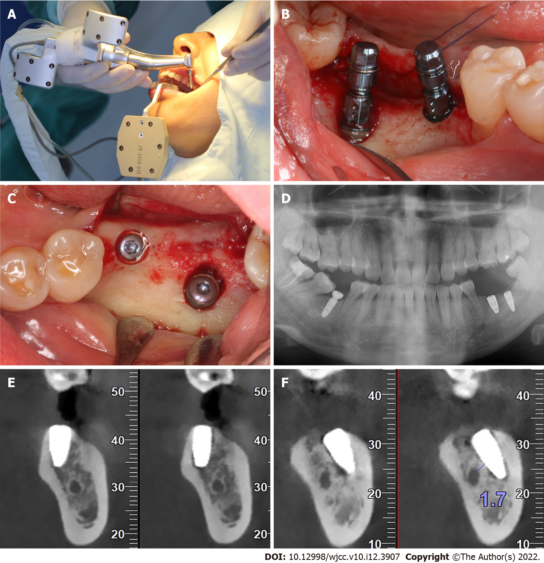
Figure 5 Implant placement surgery.
A: Surgical process; B: Buccal view of the implants; C: Occlusal view of the implant entry points; D: Panoramic radiograph after the surgery; E: Sagittal view of the first molar; F: Sagittal view of the second molar. The implant was 1.7 mm away from inferior alveolar nerve.
- Citation: Chen LW, Zhao XE, Yan Q, Xia HB, Sun Q. Dynamic navigation system-guided trans-inferior alveolar nerve implant placement in the atrophic posterior mandible: A case report. World J Clin Cases 2022; 10(12): 3907-3915
- URL: https://www.wjgnet.com/2307-8960/full/v10/i12/3907.htm
- DOI: https://dx.doi.org/10.12998/wjcc.v10.i12.3907


