Copyright
©The Author(s) 2022.
World J Clin Cases. Apr 26, 2022; 10(12): 3907-3915
Published online Apr 26, 2022. doi: 10.12998/wjcc.v10.i12.3907
Published online Apr 26, 2022. doi: 10.12998/wjcc.v10.i12.3907
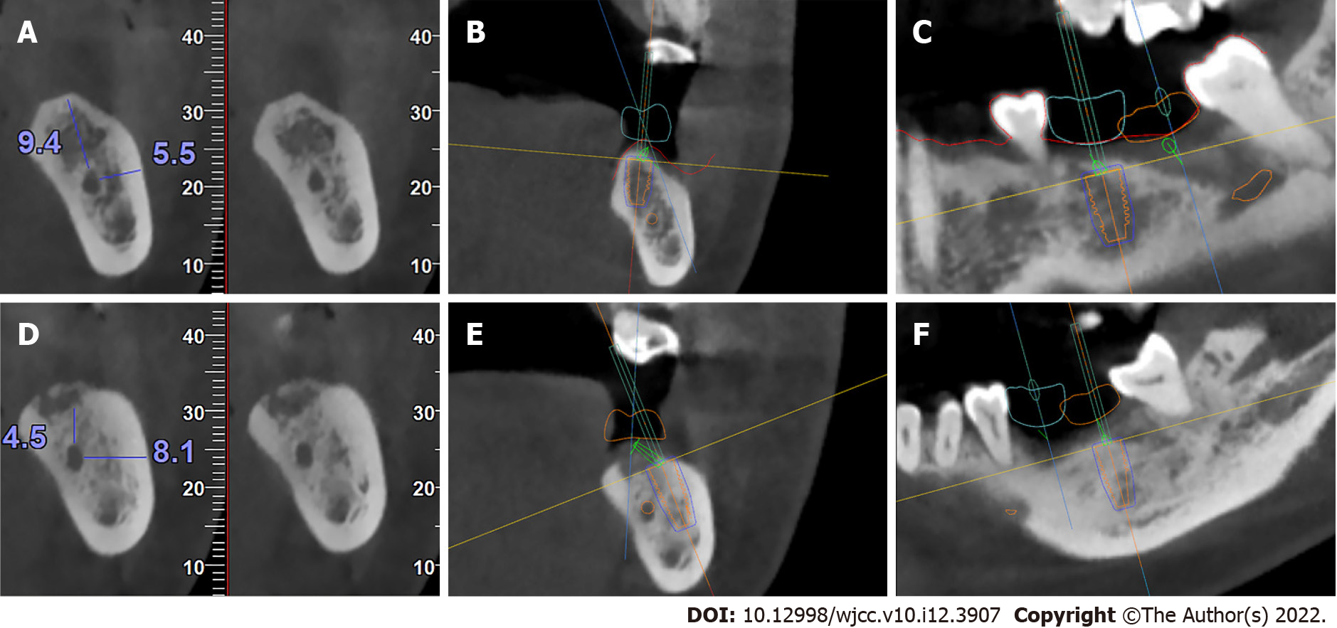
Figure 2 Arranging the proper position, length, diameter, and abutment for each implant.
A: The cone-beam computed topography (CBCT) results showed that the distance from the inferior alveolar nerve (IAN) to the crestal ridge was 9.4 mm, and the distance from the IAN to the buccal cortical bone wall was 5.5 mm in the first molar position; B: The sagittal view of the designed implant for the first molar; C: The coronal view of the designed implant for the first molar; D: The CBCT results showed that the distance from the IAN to the crestal ridge was 4.5 mm, and the distance from the IAN to the buccal cortical bone wall was 8.1 mm in the second molar position; E: The sagittal view of the designed implant for the second molar; F: The coronal view of the designed implant for the second molar.
- Citation: Chen LW, Zhao XE, Yan Q, Xia HB, Sun Q. Dynamic navigation system-guided trans-inferior alveolar nerve implant placement in the atrophic posterior mandible: A case report. World J Clin Cases 2022; 10(12): 3907-3915
- URL: https://www.wjgnet.com/2307-8960/full/v10/i12/3907.htm
- DOI: https://dx.doi.org/10.12998/wjcc.v10.i12.3907


