Copyright
©The Author(s) 2022.
World J Clin Cases. Apr 26, 2022; 10(12): 3886-3892
Published online Apr 26, 2022. doi: 10.12998/wjcc.v10.i12.3886
Published online Apr 26, 2022. doi: 10.12998/wjcc.v10.i12.3886
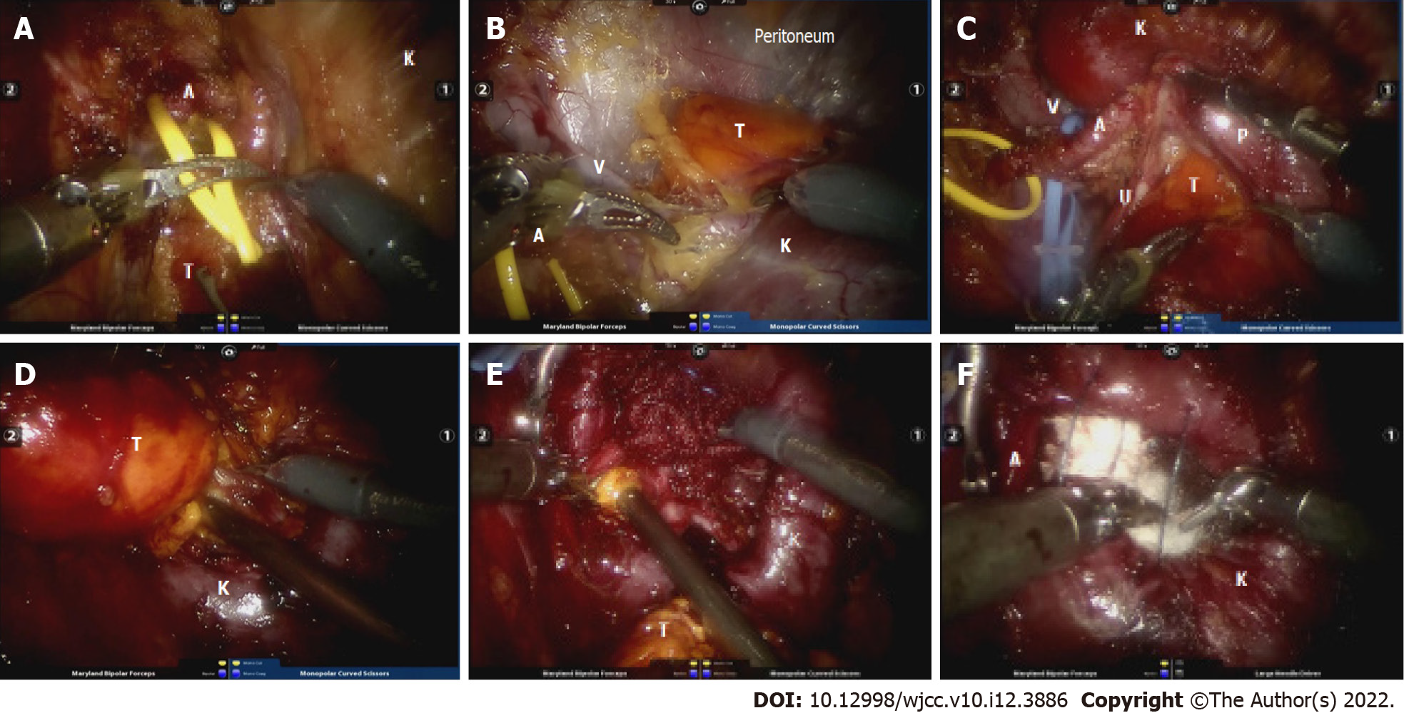
Figure 2 Surgical procedure.
A: Right renal artery (A) dissociation using the RP approach; B: The kidney (K) was thoroughly dissociated and the renal vein (V) was exposed through a good viewpoint from the dorsal to the ventral space; C: Initial tumor (T) exposure around the hilum area and ureter (U) dissociation. The artery, vein, ureter and the pelvis (P) were completely separated from the mass, respectively; D: Part of the mass was resected along its base; E: Residual tumor tissue was gradually aspirated using an aspirator; F: Pre-suture and plugging hemostatic gauzes. After pressing on the tamping for several minutes, the defect was tightly closed.
- Citation: Luo SH, Zeng QS, Chen JX, Huang B, Wang ZR, Li WJ, Yang Y, Chen LW. Successful robot-assisted partial nephrectomy for giant renal hilum angiomyolipoma through the retroperitoneal approach: A case report. World J Clin Cases 2022; 10(12): 3886-3892
- URL: https://www.wjgnet.com/2307-8960/full/v10/i12/3886.htm
- DOI: https://dx.doi.org/10.12998/wjcc.v10.i12.3886


