Copyright
©The Author(s) 2022.
World J Clin Cases. Apr 26, 2022; 10(12): 3856-3865
Published online Apr 26, 2022. doi: 10.12998/wjcc.v10.i12.3856
Published online Apr 26, 2022. doi: 10.12998/wjcc.v10.i12.3856
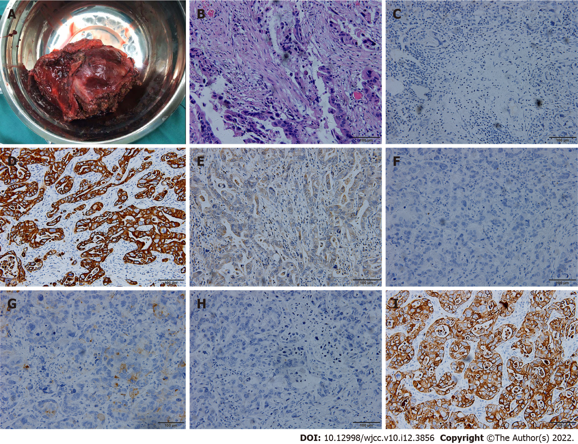
Figure 8 Surgical specimen, hematoxylin-eosin staining and immunohistochemical examination.
A: Surgical specimen; B: Hematoxylin-eosin staining showed tumor cells grew in infiltrating glandular ducts and nests (× 200); C: Immunohistochemical examination indicated ARGINASE-1 (-) (× 200); D: Immunohistochemical examination indicated CK19 (+) (× 200); E: Immunohistochemical examination indicated GPC-3 (partial +) (× 200); F: Immunohistochemical examination indicated hep-par (-) (× 200); G: Immunohistochemical examination indicated CEA (partial+) (× 200); H: Immunohistochemical examination indicated CK20 (-) (× 200); I: Immunohistochemical examination indicated CK7 (+) (× 200).
- Citation: Zhang B, Li S, Liu ZY, Peiris KGK, Song LF, Liu MC, Luo P, Shang D, Bi W. Successful multimodality treatment of metastatic gallbladder cancer: A case report and review of literature. World J Clin Cases 2022; 10(12): 3856-3865
- URL: https://www.wjgnet.com/2307-8960/full/v10/i12/3856.htm
- DOI: https://dx.doi.org/10.12998/wjcc.v10.i12.3856


