Copyright
©The Author(s) 2022.
World J Clin Cases. Apr 26, 2022; 10(12): 3842-3848
Published online Apr 26, 2022. doi: 10.12998/wjcc.v10.i12.3842
Published online Apr 26, 2022. doi: 10.12998/wjcc.v10.i12.3842
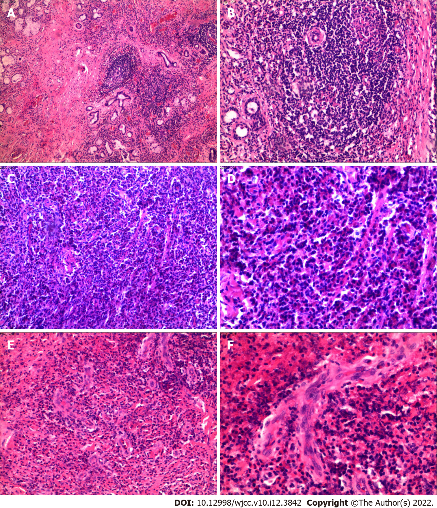
Figure 3 No evidence of malignancy was found.
Final histopathologic examination diagnosed angiomatosis with inflammatory cells. A and B: Lymphoid follicle formation (A: Perspective picture; B: Partial picture); C and D: Eosinophilic abscess area (C: Perspective picture; D: Partial picture); E and F: Obvious vascular hyperplasia (E: Perspective picture; F: Partial picture). B, D, and F are multiples higher than A, C, and E from the same area, respectively. The observed eosinophilic inflammation led to the final diagnosis of Kimura’s disease.
- Citation: Li W. Kimura's disease in soft palate with clinical and histopathological presentation: A case report. World J Clin Cases 2022; 10(12): 3842-3848
- URL: https://www.wjgnet.com/2307-8960/full/v10/i12/3842.htm
- DOI: https://dx.doi.org/10.12998/wjcc.v10.i12.3842


